Анатомия человека: атлас. Билич Г.Л., Крыжановский В.А. - Том 2. Внутренние органы. В 3-х томах.
|
|
|
|
ЛИМФОИДНАЯ СИСТЕМА (ОРГАНЫ КРОВЕТВОРЕНИЯ И ИММУННОЙ СИСТЕМЫ)
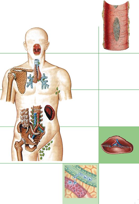 Таблица 25. Лимфоидная система
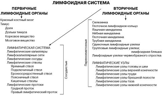
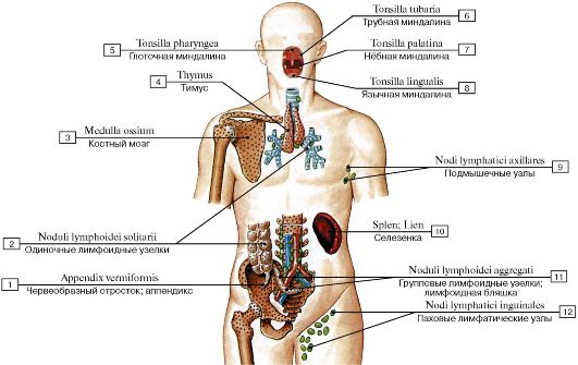 Рис. 482. Расположение органов иммунной системы (схема):
1 - Appendix; Vermiform appendix; 2 - Solitary lymphoid nodules; 3 - Bone marrow; 4 - Thymus; 5 - Pharyngeal tonsil; 6 - Tubal tonsil; 7 - Palatine tonsil; 8 - Lingual tonsil; 9 - Axillary lymph nodes; 10 - Spleen; 11 - Aggregated lymphoid nodules; 12 - Inguinal lymph
nodes
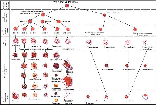 Рис 483. Постэмбриональный гемоцитопоэз и лимфоцитопоэз (схема):
I - VI - стадии дифференцировки клеток и клеток лимфоидного ряда: I - IV- морфологически неидентифицируемые клетки; V - VI - морфологически идентифицируемые клетки; КОЕ - колониеобразующие единицы; Г - гранулоциты; Μ - моноциты; Э - эритроциты; МГЦ - мегакариоцит; Эо - эозинофил; Гн - гранулярный
нейтрофил (по Н.А. Юриной)
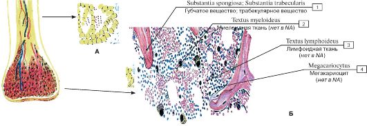 Рис. 484. Костный мозг (А - желтый, Б - красный):
1 - Spongy bone; Trabecular bone; 2 - Myeloid tissue; 3 - Lymphoid tissue; 4 - Megacaryocyte; Thromboblastus
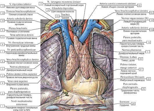 Рис. 485. Топография тимуса:
1 - Anterior mediastinal nodes; 2 - Parietal pleura, diaphragmatic part; 4 - Superior vena cava; 3 - Right lung, superior lobe; upper lobe; 5 - Phrenic nerve; 6 - Right brachiocephalic vein; 7 - Pericardiacophrenic veins; 8 - Internal thoracic veins; 11 - Right subclavian vein; 9 - Brachial plexus; 10 - Right subclavian artery; 12 - Brachiocephalic trunk; 13 - Inferior thyroid vein; 17 - Left recurrent laryngeal nerve; 14 - Thyroid gland; 15 - Trachea; 16 - Left common carotid artery; 6a - Left brachiocephalic vein; 18 - Left vagus nerve [X]; 10a - Left subclavian artery; 19 - Parietal pleura, mediastinal part; 20 - Thymus, lobe; 21 - Left lung, anterior border; 22 - Fibrous pericardium;
23 - Sternal part; 24 - Sternum
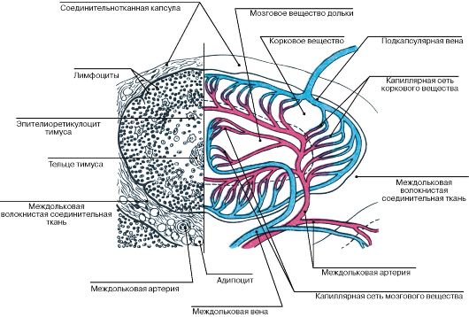 Рис. 486. Долька тимуса, строение и кровоснабжение (схема)
(рис. Ю.И. Афанасьева)
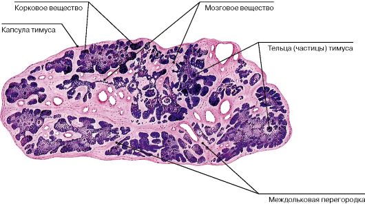 Рис. 487. Строение тимуса, гистологический срез
 Рис. 488. Тимические тельца в мозговом веществе тимуса
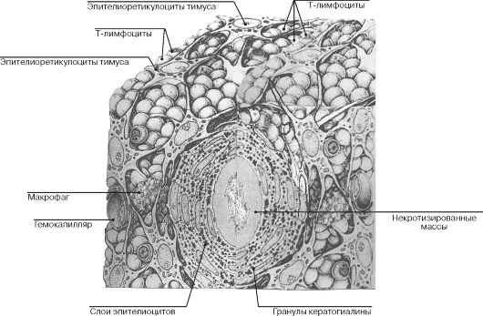 Рис. 489. Ультрамикроскопическое строение тимуса
(по Р. Крстичу, с изменениями)
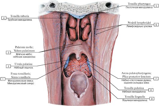 Рис. 490. Лимфоидные органы глотки, фронтальный разрез глотки, вид изнутри:
1 - Tonsillar sinus; Tonsillar fossa; Tonsillar bed; 2 - Uvula; 3 - Soft palate; 4 - Tubal tonsil; 5 - Pharyngeal tonsil; 6 - Lymphoid nodules; 7 - Palatopharyngeal arch; Posterior pillar of fauces; 8 - Palatine tonsil; 9 - Lingual tonsil
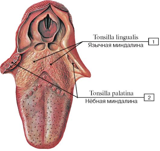 Рис. 491. Язычная и глоточная миндалины:
1 - Lingual tonsil; 2 - Palatine tonsil
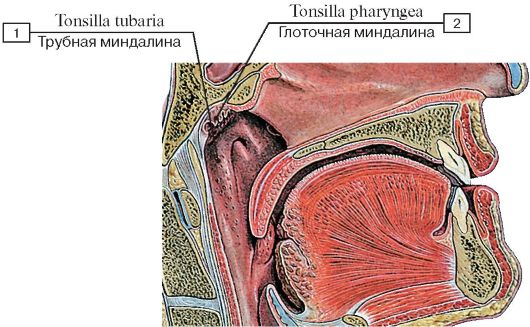 Рис. 492. Трубная и глоточная миндалины:
1 - Tubal tonsil; 2 - Pharyngeal tonsil
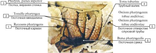 Рис. 493. Глоточная миндалина, вид снизу:
1 - Pharyngeal recess; 2 - Pharyngeal tonsil; 3 - Pharynx, roof; 4 - Torus tubarius; 5 - Pharyngeal opening of auditory tube; 6 - Pharyngeal bursa
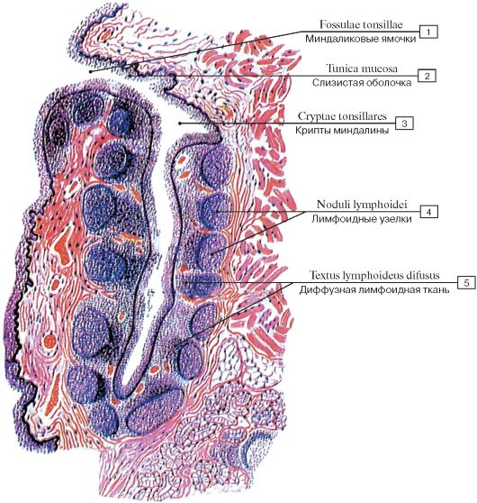 Рис. 494. Фрагмент нёбной миндалины (схема):
1 - Mucosa; Mucous membrane;
2 - Tonsillar pits; 3 - Tonsillar crypts; 4 - Lymphoid nodules;
5 - Diffuse lymphoid tissue
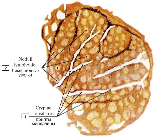 Рис. 495. Нёбная миндалина, фронтальный разрез:
1 - Tonsillar crypts; 2 - Lymphoid nodules
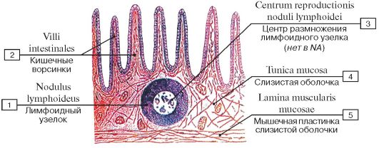 Рис. 496. Расположение лимфоидного узелка в кишечной стенке (схема):
1 - Lymphoid nodule; 2 - Villi intestinales; 3 - Center for reproduction of lymphoid nodule; 4 - Mucosa; Mucous membrane; 5 - Muscu-
laris mucosae
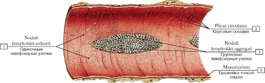 Рис. 497. Лимфоидная бляшка:
1 - Solitary lymphoid nodules; 2 - Circular folds; 3 - Aggregated lymphoid nodules; 4 - Mesentery
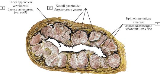 Рис. 498. Лимфоидные узелки аппендикса, поперечный разрез аппендикса:
1 - Wall of appendix; 2 - Lymphoid nodules; 3 - Epithelium mucosal tunica
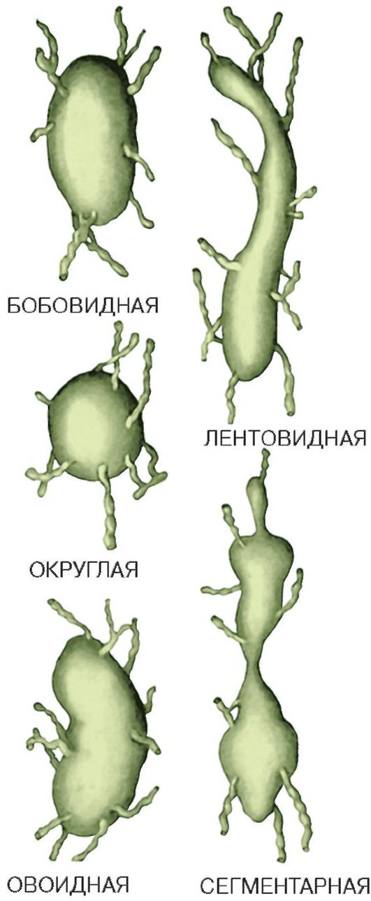 Рис. 499. Формы лимфатических узлов
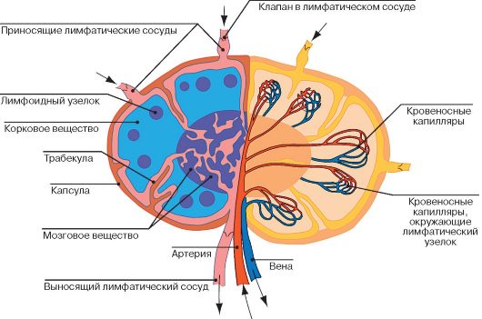 Рис. 500. Строение лимфатического узла (слева - ток лимфы, справа - ток крови)
(по Л.К. Жункейра, Ж. Карнейро)
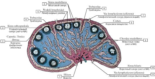 Рис. 501. Строение лимфатического узла, продольный разрез (схема):
1 - Hilum; 2 - Fibrous capsule; 3 - Subcapsular sinus; 4 - Trabeculae; 5 - Lymphoid nodules; 6 - Medullary sinus; 7 - Lymphatic vessel (afferent); 8 - Cortex; 9 - Medullary for chorda; 10 - Sinus hilaris; 11 - Lymphatic vessel (efferent)
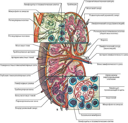 Рис. 502. Строение лимфатического узла (А - краевой синус, Б - посткапиллярная венула) (схема)
(рис. Ю.И. Афанасьева)
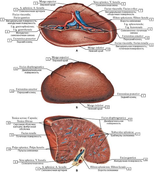 Рис.
503. Селезенка (А - вид с вентральной стороны, Б - вид с
верхнелатеральной стороны, В - вид с верхнемедиальной стороны,
поперечный разрез через ворота):
Рис.
503. Селезенка (А - вид с вентральной стороны, Б - вид с
верхнелатеральной стороны, В - вид с верхнемедиальной стороны,
поперечный разрез через ворота):
1 - Posterior extremity; 2 - Gastrosplenic ligament; 3 - Visceral surface; Gastric surface; 4 - Splenic artery; 5 - Superior border; 6 - Splenic vein; 7 - Visceral surface; Colic surface; 8 - Splenic hilum; 9 - Splenorenal ligament; Lienorenal ligament; 10 - Anterior extremity; 11 - Visceral surface; Renal surface; 12 - Inferior border; 13 - Diaphragmatic surface; 14 - Splenic pulp; 15 - Renal surface; 16 - Serosa; Serous coat; Fibrous capsule; 17 - Splenic trabeculae; 18 - Gastric surface
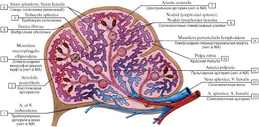 Рис. 504. Лимфоидные образования селезенки и их взаимоотношение с кровеносными сосудами (схема):
1 - Trabecular artery and vein; 2 - Penicillar arteriole; 3 - Ellipsoid macrophagal clutch; 4 - Fibrous capsule; 5 - Splenic trabecula; 6 - Splenic sinus; 7 - Central artery; 8 - Splenic lymphoid nodules; 9 - Lymphoid periarterial clutch; 10 - Red pulp; 11 - Pulpar artery;
12 - Splenic vein; 13 - Splenic artery
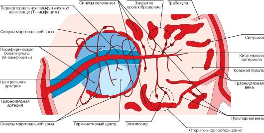 Рис. 505. Строение и кровообращение селезенки (схема)
(по Л.К. Жункейра, Ж. Карнейро)
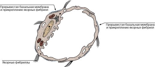 Рис. 506. Ультраструктура лимфатического капилляра (схема)
(по Л.К. Жункейра, Ж. Карнейро)
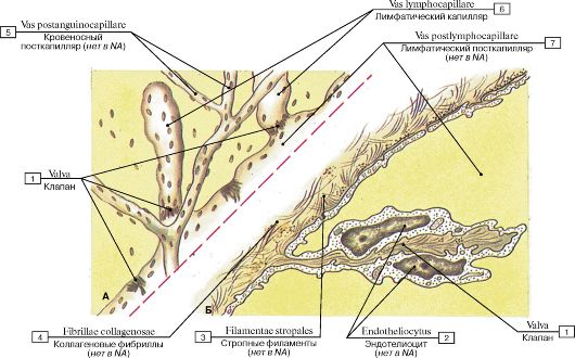 Рис.
507. Лимфатические капилляры (А - лимфатический капилляр; тотальный
препарат брюшины (импрегнация солями серебра), Б - ультраструктура
стенки лимфатических микрососудов):
Рис.
507. Лимфатические капилляры (А - лимфатический капилляр; тотальный
препарат брюшины (импрегнация солями серебра), Б - ультраструктура
стенки лимфатических микрососудов):
1 - Valve; 2 - Endotheliocytus; 3 - Stropal filaments; 4 - Collagenose fibrils; 5 - Blood postcapillary; 6 - Lymphatic capillary; 7 - Lymphatic postcapillary (по О.В. Волковой)
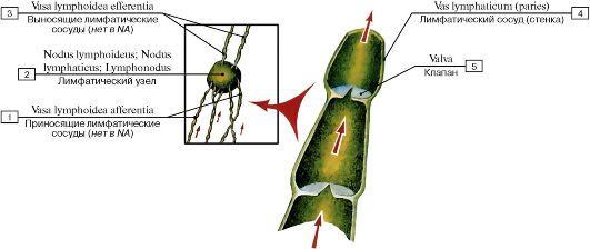 Рис. 508. Строение лимфатических сосудов (схема):
1 - Afferent lymph vessels; 2 - Lymph node; 3 - Efferent lymph vessels; 4 - Lymphatic vessel (Wall); 5 - Valve
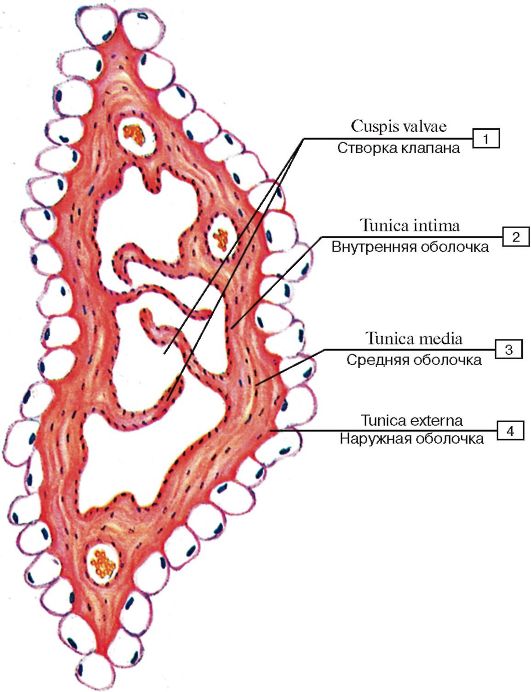 Рис. 509. Магистральный лимфатический сосуд мышечного типа:
1 - Cusp valve; 2 - Tunica intima; 3 - Tunica media; 4 - Tunica externa (по О.В. Волковой)
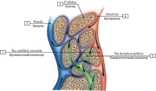 Рис. 510. Отношение между кровеносными и лимфатическими капиллярами (схема):
1 - Blood capillary; 2 - Venule; 3 - Cells; 4 - Arteriole; 5 - Lymphatic capillary
 Рис. 511. Миграция лейкоцитов через кровеносные и лимфатические сосуды (схема):
1 - Connective tissue; 2 - Leukocyt; 3 - Lymphatic capillary; 4 - Venule
Таблица 26. Характеристика лимфатических протоков и стволов
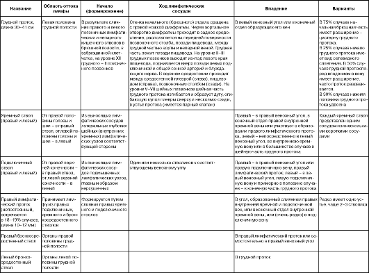 Таблица 27. Группы лимфатических узлов, к которым оттекает лимфа от некоторых внутренних органов
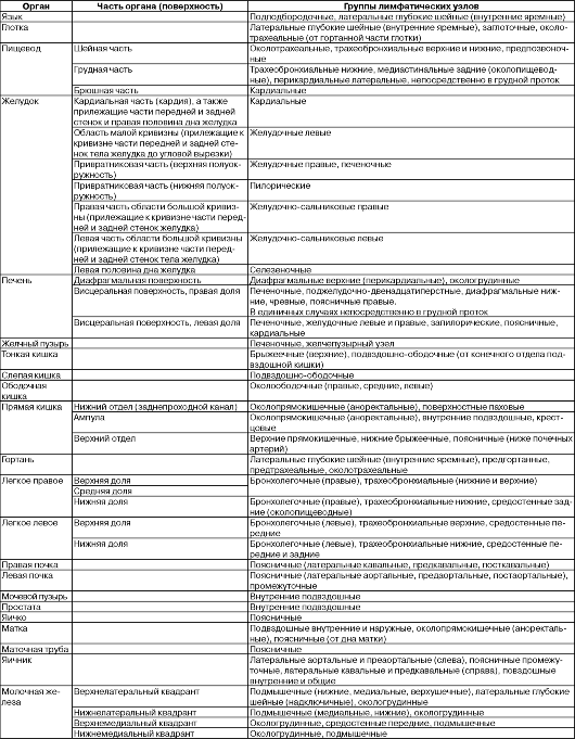
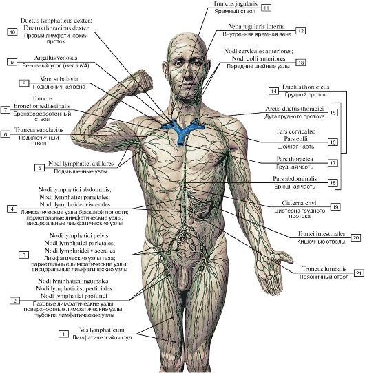 Рис. 512. Строение лимфатической системы (схема):
1 - Lymphatic vessel; 2 - Inguinal lymph nodes; Superficial lymph nodes; Deep lymph nodes; 3 - Pelvic lymph nodes; Parietal nodes; Visceral lymph nodes; 4 - Abdominal lymph nodes; Parietal nodes; Visceral lymph nodes; 5 - Axillary lymph nodes; 6 - Subclavian trunk; 7 - Bronchomediastinal trunk; 8 - Subclavian vein; 9 - Venous angle; 10 - Right lymphatic duct; Right thoracic duct; 11 - Jugular trunk; 12 - Internal jugular vein; 13 - Anterior cervical nodes; 14 = 15 + 16 + 17 + 18 - Thoracic duct; 15 - Arch of thoracic duct; 16 - Cervical part; 17 - Thoracic part; 18 - Abdominal part; 19 - Cisterna chyli; Chyle cistern; 20 - Intestinal trunks; 21 - Lumbar trunk
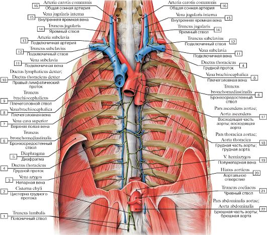 Рис. 513. Лимфатические протоки, вид спереди:
I - Lumbar trunk; 2 - Cisterna chyli; Chyle cistern; 3 - Azygos vein; 4 - Thoracic duct; 5 - Diaphragm; 6 - Bronchomediastinal trunk; 7 - Superior vena cava; 8 - Brachiocephalic vein; 9 - Brachiocephalic trunk; 10 - Right lymphatic duct; Right thoracic duct;
II - Subclavian vein; 12 - Subclavian trunk; 13 - Subclavian artery; 14 - Jugular trunk; 15 - Internal jugular vein; 16 - Common carotid artery; 17 - Ascending aorta; 18 - Thoracic aorta; 19 - Hemi-azygos vein; Inferior hemi-azygos vein; 20 - Aortic hiatus; 21 - Coeliac trunk;
22 - Abdominal aorta
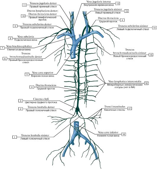 Рис. 514. Лимфатические протоки (схема):
1 - Left lumbar trunk; 2 - Right lumbar trunk; 3 - Cisterna chyli; Chyle cistern; 4 - Thoracic duct; 5 - Superior vena cava; 6 - Right bronchomediastinal trunk; 7 - Brachiocephalic vein; 8 - Subclavian vein; 9 - Right subclavian trunk; 10 - Right lymphatic duct; Right thoracic duct; 11 - Right jugular trunk; 12 - Internal jugular vein; 13 - Left jugular trunk; 14 - Left subclavian trunk; 15 - Left bronchomediastinal trunk; 16 - Intercostal lymphatic vessel; 17 - Intestinal trunks; 18 - Inferior vena cava
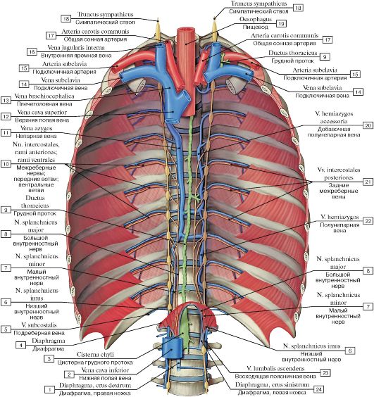 Рис. 515. Грудной проток, вид спереди:
1 - Diaphragm, right crus; 2 - Inferior vena cava; 3 - Cisterna chyli; Chyle cistern; 4 - Diaphragm; 5 - Subcostal vein; 6 - Least splanchnic nerve; Lowest splanchnic nerve; 7 - Lesser splanchnic nerve; 8 - Greater splanchnic nerve; 9 - Thoracic duct; 10 - Intercostal nerves, anterior rami; ventral rami; 11 - Azygos vein; 12 - Superior vena cava; 13 - Brachiocephalic vein; 14 - Subclavian vein; 15 - Subclavian artery; 16 - Internal jugular vein; 17 - Common carotid artery; 18 - Sympathetic trunk; 19 - Oesophagus; 20 - Accessory hemi-azygos vein; Superior hemi-azygos vein; 21 - Posterior intercostal veins; 22 - Hemi-azygos vein; Inferior hemi-azygos vein; 23 - Ascending lumbar vein;
24 - Diaphragm, left crus
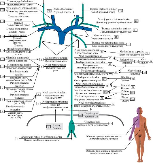 Рис. 516. Отток лимфы от различных областей тела в правый лимфатический и грудной протоки (схема):
I - Abdomen; Pelvis; Lower limb; 2 - Liver; 3 - Superior diaphragmatic nodes; 4 - Paravertebral nodes; 5 - Parasternal nodes; 6 - Paramammary nodes; 7 - Posterior intercostal part; 8 - Posterior thoracic wall; 9 - Anterior thoracic wall; 10 - Anterior intercostal part;
II - Anterior mediastinum; 12 - Breast; 13 - Bronchomediastinal trunk; 14 - Right lymphatic duct; Right thoracic duct; 15 - Right subclavian trunk; 16 - Right internal jugular vein; 17 - Right jugular trunk; 18 - Thoracic duct; 19 - Left jugular trunk; 20 - Left internal jugular vein; 21 - Left subclavian trunk; 22 - Subclavian vein; 23 - Brachiocephalic nodes; 24 - Prepericardial nodes; 25 - Lateral pericardial nodes; 26 - Paraesophageal nodes; 27 - Paratracheal nodes; 28 - Tracheobronchial nodes; 29 - Bronchopulmonary nodes; 30 - Intrapulmonary nodes; 31 - Superior diaphragmatic nodes; 32 - Superior mediastinum; 33 - Pericardium; 34 - Heart; 35 - Oesophagus; 36 - Trachea; 37 - Bronchi; 38 - Lung; 39 - Pleura; 40 - Diaphragm; 41 - Cisterna chyli; Chyle cistern
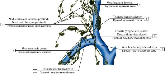 Рис. 517. Правый венозный угол и впадающие в него правый яремный ствол, правый подключичный ствол и правый
лимфатический проток (схема):
1 - Right subclavian trunk; 2 - Right subclavian vein; 3 - Lateral cervical deep nodes; 4 - Internal jugular vein; 5 - Right jugular trunk;
6 - Right lymphatic duct; Right thoracic duct; 7 - Right brachiocephalic vein
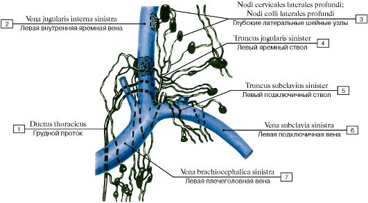 Рис. 518. Левый венозный угол и впадающие в него лимфатические стволы и грудной лимфатический проток (схема):
1 - Thoracic duct; 2 - Left internal jugular vein; 3 - Lateral cervical deep nodes; 4 - Left jugular trunk; 5 - Left subclavian trunk; 6 - Left
subclavian vein; 7 - Left brachiocephalic vein
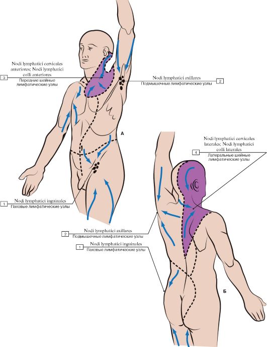 Рис. 519. Области оттока лимфы через региональные лимфатические узлы (А - вид спереди, Б - вид сзади):
1 - Inguinal lymph nodes; 2 - Axillary lymph nodes; 3 - Anterior cervical lymph nodes; 4 - Lateral cervical lymph nodes
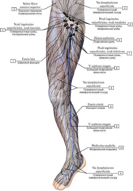 Рис. 520. Поверхностные лимфатические сосуды и узлы нижней конечности, вид спереди:
1 - Fascia lata; 2 - Superficial inguinal nodes, lateral nodes; 3 - Anterior superior iliac spine; 4 - Superficial lymph vessel; Superficial inguinal nodes, medial nodes; 6 - Saphenous opening; 7 - Superficial inguinal nodes, inferior nodes; 8 - Great saphenous vein; Long
saphenous vein; 9 - Deep fascia of leg; 10 - Medial malleolus
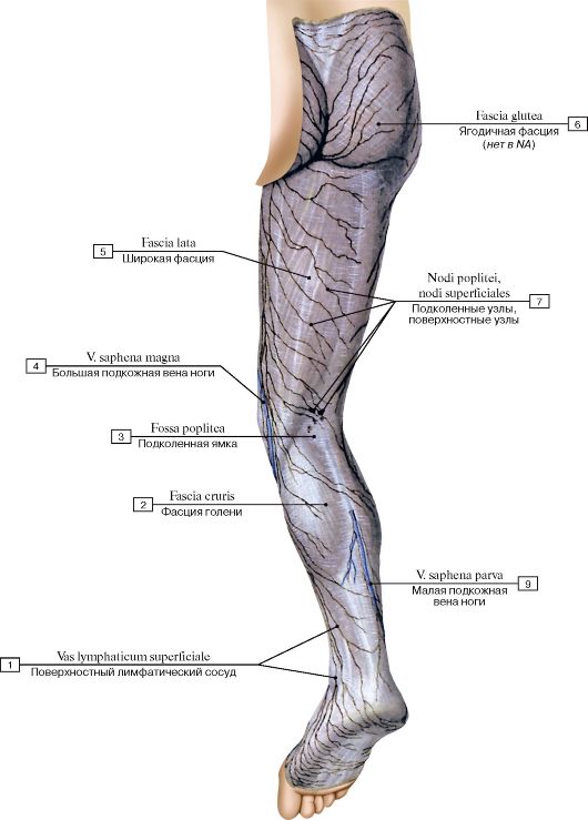 Рис. 521. Поверхностные лимфатические сосуды и узлы нижней конечности, вид сзади:
1 - Superficial lymph vessel; 2 - Deep fascia of leg; 3 - Popliteal fossa; 4 - Great saphenous vein; Long saphenous vein; 5 - Fascia lata; 6 - Fascia glutea; 7 - Popliteal nodes; Superficial nodes; 8 - Small saphenous vein; Short saphenous vein
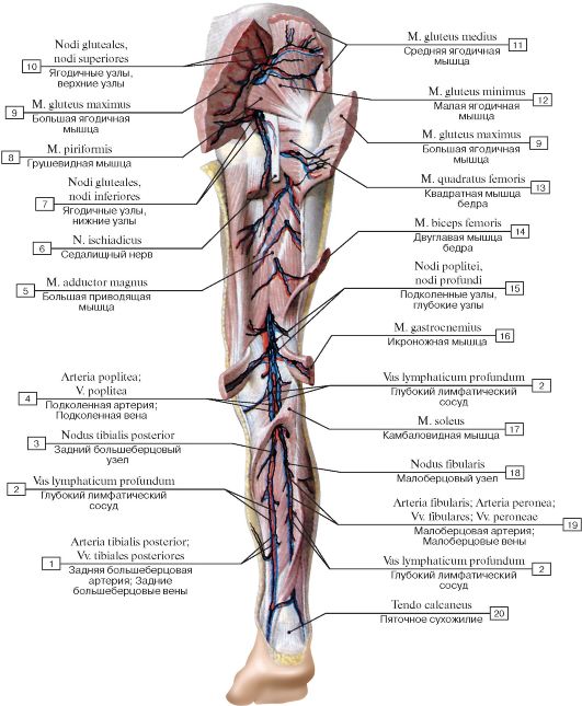 Рис. 522. Глубокие лимфатические сосуды и узлы нижней конечности, вид сзади:
1 - Posterior tibial artery; Posterior tibial veins; 2 - Deep lymph vessel; 3 - Posterior tibial node; 4 - Popliteal artery; Popliteal vein; 5 - Adductor magnus; 6 - Sciatic nerve; 7 - Gluteal nodes, inferior nodes; 8 - Piriformis; 9 - Gluteus maximus; 10 - Gluteal nodes, superior nodes; 11 - Gluteus medius; 12 - Gluteus minimus; 13 - Quadratus femoris; 14 - Biceps femoris; 15 - Popliteal nodes, deep nodes; 16 - Gastrocnemius; 17 - Soleus; 18 - Fibular node; Peroneal node; 19 - Fibular artery; Peroneal artery; Fibular veins;
Peroneal veins; 20 - Calcaneal tendon
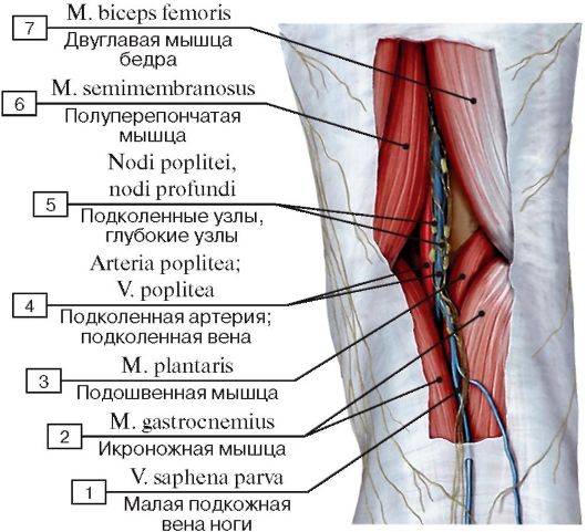 Рис. 523. Глубокие лимфатические узлы подколенной области:
1 - Small saphenous vein; Short saphenous vein; 2 - Gastrocnemius; 3 - Plantaris; 4 - Popliteal artery; Popliteal vein; 5 - Popliteal nodes; Deep nodes; 6 - Semimembranosus; 7 - Biceps femoris
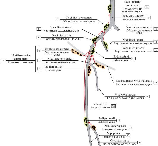 Рис. 524. Лимфатические узлы нижней конечности и таза (схема):
1 - Inferior nodes; 2 - Superomedial nodes; 3 - Superolateral nodes; 4 = 1 + 2 + 3 - Superficial nodes; 5 - External iliac nodes; 6 - External iliac vein; 7 - Common iliac nodes; 8 - Intermediate lumbar nodes; 9 - Inferior vena cava; 10 - Common iliac vein; 11 - Internal iliac nodes; 12 - Internal iliac vein; 13 - Deep nodes; 14 - Inguinal ligament; 15 - Great saphenous vein; Long saphenous vein; 16 - Femoral vein; 17 - Popliteal vein; 18 - Small saphenous vein; Short saphenous vein
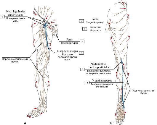 Рис.
525. Отток лимфы от нижней конечности (А - передняя поверхность, Б -
задняя поверхность) (стрелки указывают основное направление
лимфатического оттока) (схема):
Рис.
525. Отток лимфы от нижней конечности (А - передняя поверхность, Б -
задняя поверхность) (стрелки указывают основное направление
лимфатического оттока) (схема):
1 - Superficial inguinal nodes; 2 - Penis; 3 - Great saphenous vein; Long saphenous vein; 4 - Small saphenous vein; Short saphenous vein;
5 - Popliteal nodes, superficial nodes; 6 - Scrotum; 7 - Anus
Лимфа оттекает от нижних конечностей по поверхностным и глубоким лимфатическим сосудам.
Наиболее крупные из них (коллекторные) обычно следуют по ходу поверхностных (большой и малой поверхностных вен ноги) и глубоких вен (подколенной и бедренной).
Лимфатические сосуды связаны между собой анастомозами, расположенными главным образом в подколенной и паховой областях.
Поверхностные лимфатические сосуды осуществляют отток лимфы в основном от кожи и подкожной основы, глубокие - от мышц и суставов.
Поверхностные лимфатические сосуды образуют переднемедиальный и заднелатеральный пучки. Переднемедиальный пучок, проходящий вдоль большой поверхностной вены ноги к паховым лимфатическим узлам, отводит лимфу от кожи и подкожной основы кроме латерального края стопы и узкой полосы кожи на икре. От этих участков лимфа отходит по сосудам заднелатерального пучка (см. Б).
Заднелатеральный пучок проходит вдоль малой поверхностной вены ноги и сначала отводит лимфу в поверхностные подколенные лимфатические узлы, от которых она оттекает через глубокие подколенные лимфатические узлы в глубокие паховые лимфатические узлы.
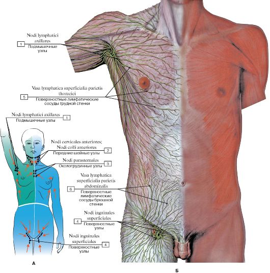 Рис.
526. Отток лимфы от передних стенок груди и живота (А - отток лимфы от
передней стенки груди, живота и верхних конечностей (схема), Б -
лимфатические сосуды передних стенок груди и живота):
Рис.
526. Отток лимфы от передних стенок груди и живота (А - отток лимфы от
передней стенки груди, живота и верхних конечностей (схема), Б -
лимфатические сосуды передних стенок груди и живота):
1 - Axillary lymph nodes; 2 - Anterior cervical nodes; 3 - Parasternal nodes; 4 - Superficial inguinal nodes; 5 - Superficial lymphatic vessels
of thoracic wall; 6 - Superficial lymphatic vessels of abdominal wall
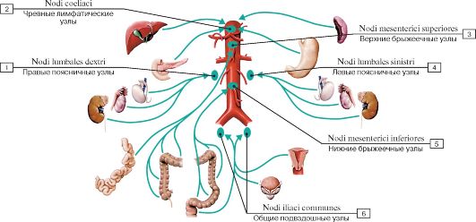 Рис. 527. Группы лимфатических узлов полости таза и брюшной полости, стрелками показан отток лимфы (схема):
1 - Right lumbar nodes; 2 - Coeliac nodes; 3 - Superior mesenteric nodes; 4 - Left lumbar nodes; 5 - Inferior mesenteric nodes; 6 - Common iliac nodes
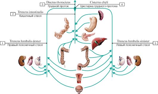 Рис. 528. Лимфатические стволы полости таза и брюшной полости, стрелками показан отток лимфы (схема):
1 - Right lumbar trunk; 2 - Intestinal trunk; 3 - Thoracic duct; 4 - Cisterna chyli; Chyle cistern; 5 - Left lumbar trunk
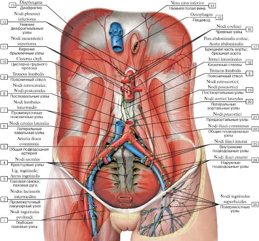 Рис. 529. Паховые, подвздошные и поясничные лимфатические узлы:
1 - Deep inguinal nodes; 2 - Intermediate lacunar node; 3 - Inguinal ligament; 4 - Sacral nodes; 5 - Common iliac artery; 6 - Lateral caval nodes; 7 - Intermediate lumbar nodes; 8 - Postcaval nodes; 9 - Lumbar trunk; 10 - Cisterna chyli; Chyle cistern; 11 - Superior mesenteric nodes; 12 - Inferior diaphragmatic nodes; 13 - Diaphragm; 14 - Inferior vena cava; 15 - Oesophagus; 16 - Coeliac nodes; 17 - Abdominal aorta; 18 - Intestinal trunks; 19 - Postaortic nodes; 20 - Lateral aortic nodes; 21 -Pre-aortic nodes; 22 - Common iliac nodes; 23 - Internal
iliac nodes; 24 - External iliac nodes; 25 - Superficial inguinal nodes
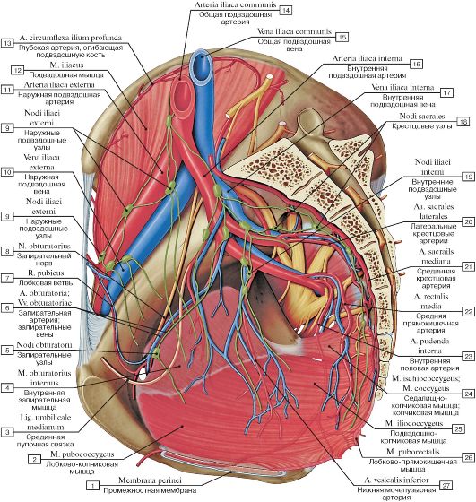 Рис. 530. Лимфатические сосуды и узлы таза, сагиттальный разрез, вид изнутри, слева:
1 - Perineal membrane; 2 - Pubococcygeus; 3 - Median umbilical ligament; 4 - Obturator internus; 5 - Obturator nodes; 6 - Obturator artery; Obturator veins; 7 - Pubic branch; 8 - Obturator nerve; 9 - External iliac nodes; 10 - External iliac vein; 11 - External iliac artery; 12 - Iliacus; 13 - Deep circumflex iliac artery; 14 - Common iliac artery; 15 - Common iliac vein; 16 - Internal iliac artery; 17 - Internal iliac vein; 18 - Sacral nodes; 19 - Internal iliac nodes; 20 - Lateral sacral arteries; 21 - Median sacral artery; 22 - Middle rectal artery; 23 - Internal pudendal artery; 24 - Ischiococcygeus; Coccygeus; 25 - Iliococcygeus; 26 - Puborectalis; 27 - Inferior vesical artery
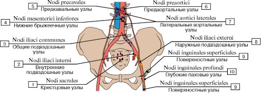 Рис. 531. Расположение тазовых лимфатических узлов (схема):
1 - Sacral nodes; 2 - Internal iliac nodes; 3 - Common iliac nodes; 4 - Inferior mesenteric nodes; 5 - Precaval nodes; 6 - Pre-aortic nodes; 7 - Lateral aortic nodes; 8 - External iliac nodes; 9 - Superficial inguinal nodes; 10 - Deep inguinal nodes
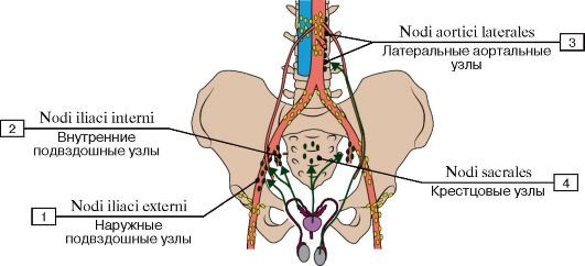 Рис. 532. Отток лимфы от мужских половых органов (схема):
1 - External iliac nodes; 2 - Internal iliac nodes; 3 - Lateral aortic nodes; 4 - Sacral nodes
 Рис. 533. Отток лимфы от мочевого пузыря и мочеиспускательного канала (схема):
1 - Inguinal lymph nodes, superficial inguinal nodes; 2 - Internal iliac nodes; 3 - Common iliac nodes; 4 - External iliac nodes
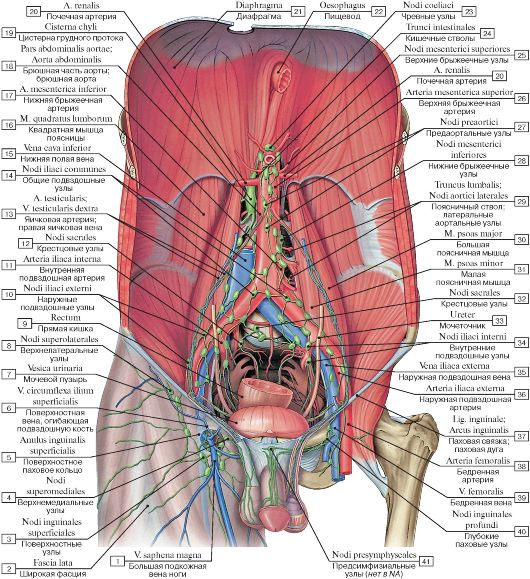 Рис. 534. Лимфатические узлы таза и брюшной полости мужчины:
I - Great saphenous vein; Long saphenous vein; 2 - Fascia lata; 3 - Superficial inguinal nodes; 4 - Superomedial nodes; 5 - Superficial inguinal ring; 6 - Superficial circumflex iliac vein; 7 - Urinary bladder; 8 - Superolateral nodes; 9 - Rectum; 10 - External iliac nodes;
II - Internal iliac artery; 12 - Sacral nodes; 13 - Testicular artery; Right testicular vein; 14 - Common iliac nodes; 15 - Inferior vena cava; 16 - Quadratus lumborum; 17 - Inferior mesenteric artery; 18 - Abdominal aorta; 19 - Cisterna chyli; Chyle cistern; 20 - Renal artery; 21 - Diaphragm; 22 - Oesophagus; 23 - Coeliac nodes; 24 - Intestinal trunks; 25 - Superior mesenteric nodes; 26 - Superior mesenteric artery; 27 - Pre-aortic nodes; 28 - Inferior mesenteric nodes; 29 - Lumbar trunk; Lateral aortic nodes; 30 - Psoas major; 31 - Psoas minor; 32 - Sacral nodes; 33 - Ureter; 34 - Internal iliac nodes; 35 - External iliac vein; 36 - External iliac artery; 37 - Inguinal ligament;
38 - Femoral artery; 39 - Femoral vein; 40 - Deep inguinal nodes; 41 - Presymphyseal nodes
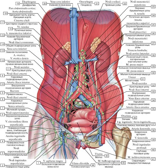 Рис. 535. Лимфатические узлы таза и брюшной полости женщины:
1 - Great saphenous vein; Long saphenous vein; 2 - Superficial inguinal nodes; 3 - Superomedial nodes; 4 - Superficial inguinal ring; 5 - Superficial circumflex iliac vein; 6 - Superolateral nodes; 7 - Urinary bladder; 8 - External iliac nodes; 9 - Rectum; 10 - Internal iliac artery; 11 - Sacral nodes; 12 - Ovarian artery; Right ovarian vein; 13 - Inferior vena cava; 14 - Common iliac nodes; 15 - Inferior mesenteric artery; 16 - Renal veins; 17 - Cisterna chyli; Chyle cistern; 18 - Abdominal aorta; 19 - Diaphragm; 20 - Inferior vena cava; 21 - Oesophagus; 22 - Coeliac nodes; 23 - Superior mesenteric nodes; 24 - Renal artery; 25 - Superior mesenteric artery; 26 - Pre-aortic nodes; 27 - Inferior mesenteric nodes; 28 - Lumbar trunk; Lateral aortic nodes; 29 - Psoas major; 30 - Psoas minor; 31 - Ureter; 32 - Sacral nodes; 33 - Internal iliac nodes; 34 - External iliac vein; 35 - External iliac artery; 36 - Ovary; 37 - Inguinal ligament; 38 - Femoral artery; 39 - Femoral vein; 40 - Deep inguinal nodes; 41 - Ischiocavernosus; 42 - Uterus; 43 - Bulbospongiosus; 44 - Glans of clitoris
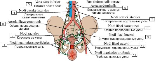 Рис. 536. Отток лимфы от женских половых органов (схема):
1 - Superficial inguinal nodes; 2 - Sacral nodes; 3 - Common iliac artery; 4 - Lateral caval nodes; 5 - Inferior vena cava; 6 - Abdominal aorta; 7 - Lateral aortic nodes; 8 - Common iliac nodes; 9 - Internal iliac nodes; 10 - External iliac nodes; 11 - Deep inguinal nodes
От наружных и внутренних половых органов лимфа оттекает в различные группы париетальных лимфатических узлов, от которых лимфа направляется в поясничные лимфатические узлы, расположенные около брюшной части аорты и нижней полой вены.
От наружных половых органов (и нижних отделов влагалища) лимфа оттекает в поверхностные и глубокие паховые лимфатические узлы, а также дополнительно (не показано) непосредственно в подвздошные лимфатические узлы.
От яичников, дна матки и в основном дистальных частей фаллопиевых трубы - длинный путь оттока лимфы
в поясничные лимфатические узлы, расположенные вокруг брюшной части аорты и нижней полой вены.
От дна и тела матки и проксимальных частей фаллопиевых труб лимфа оттекает в крестцовые, внутренние и наружные подвздошные лимфатические узлы.
От шейки матки, а также от средней и верхней частей влагалища лимфа оттекает в глубокие паховые лимфатические узлы.
Малые висцеральные лимфатические узлы матки и влагалища (околоматочные и околовлагалищные узлы, здесь не показаны) находятся в соединительной ткани таза вблизи от указанных органов.
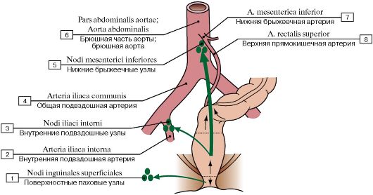 Рис. 537. Отток лимфы от прямой кишки (схема):
1 - Superficial inguinal nodes; 2 - Internal iliac artery; 3 - Internal iliac nodes; 4 - Common iliac artery; 5 - Inferior mesenteric nodes;
6 - Abdominal aorta; 7 - Inferior mesenteric artery; 8 - Superior rectal artery
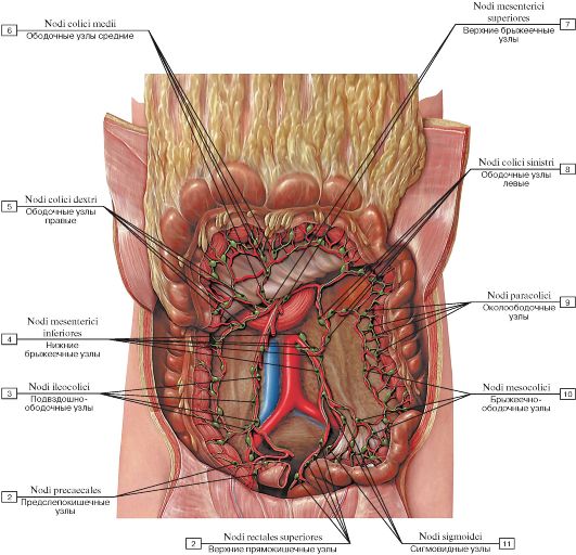 Рис. 538. Ободочно-кишечные лимфатические узлы:
1 - Superior rectal nodes; 2 - Precaecal nodes; 3 - Ileocolic nodes; 4 - Inferior mesenteric nodes; 5 - Right colic nodes; 6 - Middle colic nodes; 7 - Superior mesenteric nodes; 8 - Left colic nodes; 9 - Paracolic nodes; 10 - Mesocolic nodes; 11 - Sigmoid nodes;
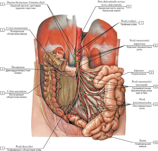 Рис. 539. Брыжеечные лимфатические узлы:
1 - Ileocolic nodes; 2 - Ascending colon; 3 - Duodenum; 4 - Transverse colon; 5 - Thoracic duct; Cisterna chyli; Chyle cistern; 6 - Abdominal aorta; 7 - Coeliac nodes; 8 - Superior mesenteric nodes; 9 - Jejunum; 10 - Intermediate mesenteric nodes; 11 - Juxta-
intestinal mesenteric nodes; 12 - Ileum
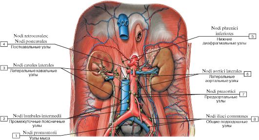 Рис. 540. Почечные, надпочечниковые и мочеточниковые лимфатические узлы:
1 - Promontorial nodes; 2 - Intermediate lumbar nodes; 3 - Lateral caval nodes; 4 - Postcaval nodes; 5 - Inferior diaphragmatic nodes;
6 - Lateral aortic nodes; 7 - Pre-aortic nodes; 8 - Common iliac nodes
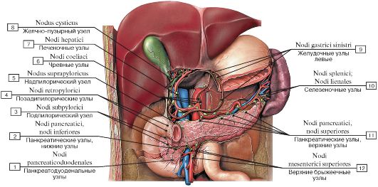 Рис. 541. Поджелудочные, селезеночные и двенадцатиперстные лимфатические узлы:
1 - Pancreaticoduodenal nodes; 2 - Pancreatic nodes, inferior nodes; 3 - Subpyloric nodes; 4 - Retropyloric nodes; 5 - Suprapyloric node; 6 - Coeliac nodes; 7 - Hepatic nodes; 8 - Cystic node; 9 - Left gastric nodes; 10 - Splenic nodes; 11 - Pancreatic nodes, superior nodes;
12 - Superior mesenteric nodes
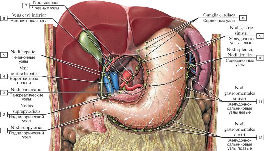 Рис. 542. Лимфатические узлы желудка, желудочно-сальниковые, печеночные и селезеночные узлы:
1 - Subpyloric nodes; 2 - Suprapyloric node; 3 - Pancreatic nodes; 4 - Hepatic portal vein; 5 - Hepatic nodes; 6 - Inferior vena cava; 7 - Coeliac nodes; 8 - Cardiac ganglia; 9 - Left gastric nodes; 10 - Splenic nodes; 11 - Left gastro-omental nodes; 12 - Right gastro-
omental nodes
Основные пути оттока лимфы от органов пищеварительной системы и селезенки (рис. 543).
Лимфа от селезенки и большинства органов пищеварительной системы оттекает непосредственно от региональных лимфатических узлов либо через вставочные («накопительные») лимфатические узлы в кишечные стволы. Исключения составляют нисходящая ободочная, сигмовидная ободочная и верхняя часть прямой кишки, от которых лимфа оттекает в левый поясничный ствол. От органов и висцеральных лимфатических узлов лимфа оттекает главным образом в три крупные «накопительные» системы:
• Чревные лимфатические узлы собирают лимфу от желудка, двенадцатиперстной кишки, селезенки, поджелудочной железы и печени. Топографически они часто не отличаются от региональных лимфатических узлов близлежащих органов верхней половины брюшной полости.
• Верхние брыжеечные узлы собирают лимфу от тощей, подвздошной, восходящей ободочной и поперечной ободочной кишок.
• Нижние брыжеечные узлы собирают лимфу от нисходящей ободочной, сигмовидной ободочной и прямой кишок.
От этих собирательных лимфатических узлов лимфа оттекает преимущественно через кишечные стволы в цистерну грудного протока. Кроме того, имеется добавочный путь оттока в цистерну грудного протока через левые поясничные лимфатические узлы.
Основные пути оттока лимфы от нижних конечностей, органов таза и забрюшинного пространства (рис. 544).
Лимфа от этих органов оттекает главным образом в правый и левый поясничные стволы. Перечисленные ниже группы лимфатических узлов являются важными для органов забрюшинного пространства, таза и нижней конечности:
• Общие подвздошные лимфатические узлы собирают лимфу от органов таза и нижней конечности.
• Правые и левые поясничные лимфатические узлы являются собирательными для общих подвздошных лимфатических узлов, а также структурно лимфатических узлов органов забрюшинного пространства и яичников (ж) и яичек (м), хотя последние и расположены в полости таза или мошонке. Так как гонады опускаются в процессе внутриутробного развития, они сохраняют свои лимфатические связи с поясничными лимфатическими узлами (аналогично с их кровоснабжением).
Подвздошные и поясничные лимфатические узлы являются париетальными, куда относятся также диафрагмальные и надчревные лимфатические узлы. Околопрямокишечные и околоматочные лимфатические узлы являются висцеральными.
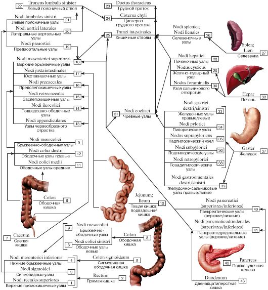 Рис. 543. Основные пути оттока лимфы от органов пищеварительной системы и селезенки (схема):
1 - Superior rectal nodes; 2 - Rectum; 3 - Sigmoid nodes; 4 - Inferior mesenteric nodes; 5 - Sigmoid colon; 6 - Left colic nodes; 7 - Caecum; 8 - Colon; 9 - Mesocolic nodes; 10 - Jejunum; Ileum; 11 - Middle colic nodes; 12 - Right colic nodes; 13 - Appendicular nodes; 14 - Ileocolic nodes; 15 - Retrocaecal nodes; 16 - Precaecal nodes; 17 - Juxta-intestinal mesenteric nodes; 18 - Superior mesenteric nodes; 19 - Pre-aortic nodes; 20 - Lateral aortic nodes; 21 - Left lumbar nodes; 22 - Left lumbar trunk; 23 - Thoracic duct; 24 - Cisterna chyli; Chyle cistern; 25 - Intestinal trunks; 26 - Splenic nodes; 27 - Spleen; 28 - Hepatic nodes; 29 - Cystic node; 30 - Node of anterior border of omental foramen; 31 - Coeliac nodes; 32 - Liver; 33 - Right/left gastric nodes; 34 - Pyloric nodes; 35 - Suprapyloric node; 36 - Subpyloric nodes; 37 - Stomach; 38 - Retropyloric nodes; 39 - Right/left gastro-omental nodes; 40 - Pancreatic nodes (superior/inferior); 41 - Pancreaticoduodenal nodes (superior/inferior); 42 - Pancreas; 43 - Duodenum
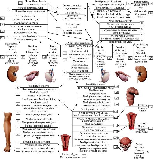 Рис. 544. Основные пути оттока лимфы от нижних конечностей, органов таза и забрюшинного пространства (схема):
1 - Uterus; Vagina; 2 - Superficial inguinal nodes; 3 - Deep inguinal nodes; 4 - Intermediate lacunar node; 5 - Medial lacunar node; 6 - Lateral lacunar node; 7 - Interiliac nodes; 8 - Lateral nodes; Medial nodes; Intermediate nodes; 9 - Obturator nodes; 10 - External iliac nodes; 11 - Promontorial nodes; 12 - Subaortic nodes; 13 - Common iliac nodes; 14 - Right kidney; Suprarenal gland; Adrenal gland; 15 - Right ovary; Uterine tube; 16 - Right testis; Epididymis; 17 - Postcaval nodes; 18 - Precaval nodes; 19 - Lateral caval nodes; 20 - Right lumbar nodes; 21 - Right lumbar trunk; 22 - Intestinal trunks; 23 - Intermediate lumbar nodes; 24 - Cisterna chyli; Chyle cistern; 25 - Thoracic duct; 26 - Inferior diaphragmatic nodes; 27 - Inferior epigastric nodes; 28 - Left lumbar trunk; 29 - Diaphragm; 30 - Abdominal part; 31 - Left lumbar nodes; 32 - Lateral aortic nodes; 33 - Pre-aortic nodes; 34 - Postaortic nodes; 35 - Left kidney; Suprarenal gland; Adrenal gland; 36 - Left ovary; Uterine tube; 37 - Left testis; Epididymis; 38 - Internal iliac nodes; 39 - Sacral nodes; 40 - Superior gluteal nodes; 41 - Inferior gluteal nodes; 42 - Rectum; 43 - Pelvic lymph nodes; 44 - Pararectal nodes; 45 - Para-uterine nodes; 46 - Paravaginal nodes; 47 - Lateral vesical nodes; 48 - Prevesical nodes; Postvesical nodes; 49 - Urinary bladder; Prostate
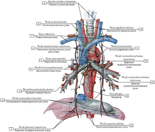 Рис. 545. Лимфатические узлы грудной полости, вид спереди:
I - Superior diaphragmatic nodes; 2 - Lateral pericardial nodes; 3 - Right main bronchus; 4 - Bronchopulmonary nodes; 5 - Superior vena cava; 6 - Anterior mediastinal nodes; 7 - Subclavian vein; 8 - Paratracheal nodes; 9 - Common carotid artery; 10 - Internal jugular vein;
II - Parasternal nodes; 12 - Superior tracheobronchial nodes; 13 - Inferior tracheobronchial nodes; 14 - Oesophagus; 15 - Prepericardial
nodes
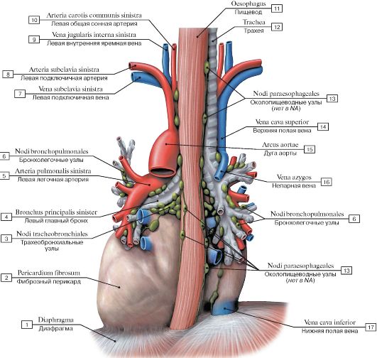 Рис. 546. Лимфатические узлы грудной полости, вид сзади:
I - Diaphragm; 2 - Fibrous pericardium; 3 - Tracheobronchial nodes; 4 - Left main bronchus; 5 - Left pulmonary artery; 6 - Bronchopulmonary nodes; 7 - Left subclavian vein; 8 - Left subclavian artery; 9 - Left internal jugular vein; 10 - Left common carotid artery;
II - Oesophagus; 12 - Trachea; 13 - Paraesophageal nodes; 14 - Superior vena cava; 15 - Arch of aorta; Aortic arch; 16 - Azygos vein;
17 - Inferior vena cava
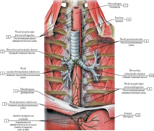 Рис. 547. Лимфатические узлы трахеи и пищевода:
1 - Cardial lymphatic ring (inconstant); 2 - Inferior diaphragmatic nodes; 3 - Diaphragm; 4 - Inferior tracheobronchial nodes; 5 - Right main bronchus; 6 - Paraesophageal lymph nodes; 7 - Oesophagus; 8 - Trachea; 9 - Paratracheal nodes; 10 - Left main bronchus;
11 - Stomach
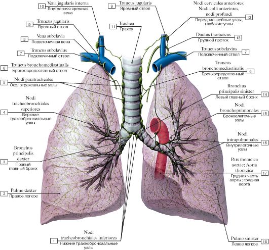 Рис. 548. Лимфатические узлы трахеи и бронхов:
1 - Inferior tracheobronchial nodes; 2 - Right lung; 3 - Right main bronchus; 4 - Superior tracheobronchial nodes; 5 - Paratracheal nodes; 6 - Bronchomediastinal trunk; 7 - Subclavian trunk; 8 - Subclavian vein; 9 - Jugular trunk; 10 - Internal jugular vein; 11 - Trachea; 12 - Anterior cervical nodes, deep nodes; 13 - Thoracic duct; 14 - Left main bronchus; 15 - Bronchopulmonary nodes; 16 - Intrapulmonary
nodes; 17 - Thoracic aorta; 18 - Left lung
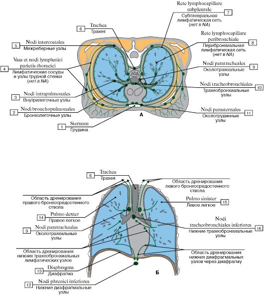 Рис.
549. Отток лимфы от трахеи и бронхов (А - поперечное сечение грудной
клетки, Б - продольное сечение грудной клетки) (схема):
Рис.
549. Отток лимфы от трахеи и бронхов (А - поперечное сечение грудной
клетки, Б - продольное сечение грудной клетки) (схема):
1 - Sternum; 2 - Bronchopulmonary nodes; 3 - Intrapulmonary nodes; 4 - Lymph nodes et vessels of thoracic wall; 5 - Intercostal nodes; 6 - Trachea; 7 - Subpleural lympatic rete; 8 - Peribronchial lympatic rete; 9 - Paratracheal nodes; 10 - Tracheobronchial nodes; 11 - Parasternal nodes; 12 - Inferior diaphragmatic nodes; 13 - Diaphragm; 14 - Right lung; 15 - Left lung; 16 - Inferior tracheobronchial nodes
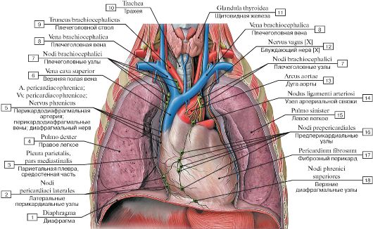 Рис. 550. Перикардиальные лимфатические узлы:
1 - Diaphragm; 2 - Lateral pericardial nodes; 3 - Parietal pleura, mediastinal part; 4 - Right lung; 5 - Pericardiacophrenic artery; Pericardiacophrenic veins; Phrenic nerve; 6 - Superior vena cava; 7 - Brachiocephalic nodes; 8 - Brachiocephalic vein; 9 - Brachiocephalic trunk; 10 - Trachea; 11 - Thyroid gland; 12 - Vagus nerve [X]; 13 - Arch ofaorta; Aortic arch; 14 - Node of ligamentum arteriosum; 15 - Left lung; 16 - Prepericardial nodes; 17 - Fibrous pericardium; 18 - Superior diaphragmatic nodes
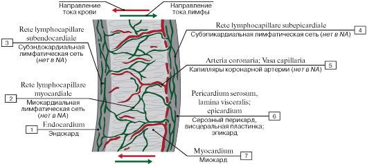 Рис. 551. Отток лимфы от миокарда (схема):
1 - Endocardium; 2 - Myocardial lymphatic rete; 3 - Subendocardial lymphatic rete; 4 - Subepicardial lymphatic rete; 5 - Capillary of coronary artery; 6 - Visceral layer; Epicardium; Serous pericardium; 7 - Myocardium
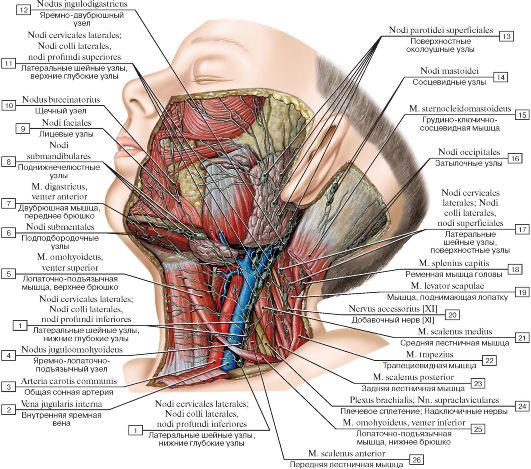 Рис. 552. Лимфатические сосуды и узлы головы и шеи:
1 - Lateral cervical nodes, inferior deep nodes; 2 - Internal jugular vein; 3 - Common carotid artery; 4 - Jugulo-omohyoid node; 5 - Omohyoid, superior belly; 6 - Submental nodes; 7 - Digastric, anterior belly; 8 - Submandibular nodes; 9 - Facial nodes; 10 - Buccinator node; 11 - Lateral cervical nodes, superior deep nodes; 12- Jugulo-digastric node; 13 - Superficial parotid nodes; 14 - Mastoid nodes; 15 - Sternocleidomastoid; 16 - Occipital nodes; 17 - Lateral cervical nodes, superficial nodes; 18 - Splenius capitis; 19 - Levator scapulae; 20 - Accessory nerve [XI]; 21 - Scalenus medius; Middle scalene; 22 - Trapezius; 23 - Scalenus posterior; Posterior scalene; 24 - Brachial plexus; Supraclavicular nerves; 25 - Omohyoid, inferior belly; 26 - Scalenus anterior; Anterior scalene
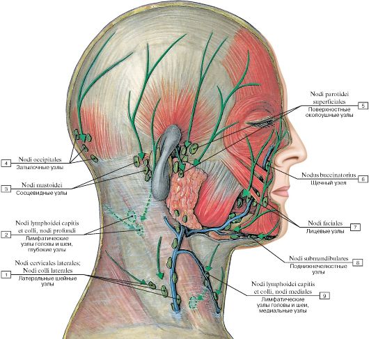 Рис. 553. Поверхностные лимфатические узлы головы, стрелками указано направление тока лимфы:
1 - Lateral cervical nodes; 2 - Lymph nodes of head and neck, deep nodes; 3 - Mastoid nodes; 4 - Occipital nodes; 5 - Superficial parotid nodes; 6 - Buccinator node; 7 - Facial nodes; 8 - Submandibular nodes; 9 - Lymph nodes of head and neck, medial nodes
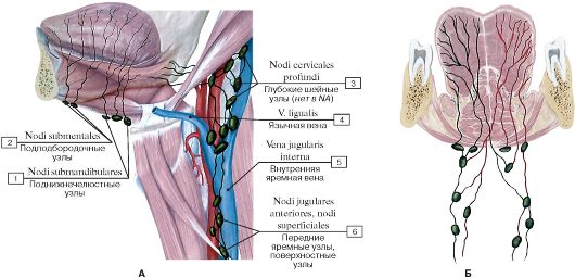 Рис. 554. Лимфатические узлы языка и полости рта (А - вид сбоку, Б - фронтальный разрез (схема)):
1 - Submandibular nodes; 2 - Submental nodes; 3 - Deep cervical nodes; 4 - Lingual vein; 5 - Internal jugular vein; 6 - Superficial nodes,
anterior jugular nodes
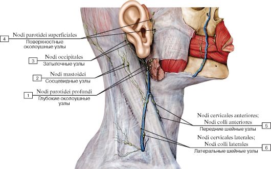 Рис. 555. Поверхностные лимфатические узлы головы и шеи, вид сбоку:
1 - Deep parotid nodes; 2 - Mastoid nodes; 3 - Occipital nodes; 4 - Superficial parotid nodes; 5 - Anterior cervical nodes; 6 - Lateral
cervical nodes
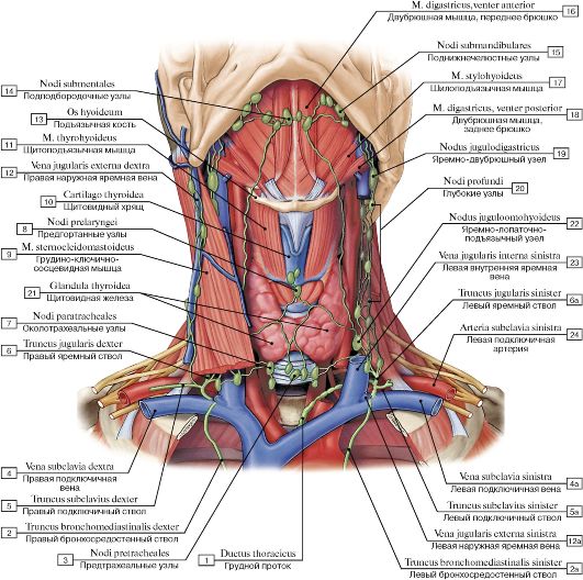 Рис. 556. Поверхностные лимфатические узлы шеи, вид спереди:
1 - Thoracic duct; 3 - Pretracheal nodes; 2 - Right bronchomediastinal trunk; 5 - Right subclavian trunk; 4 - Right subclavian vein; 6 - Right jugular trunk; 7 - Paratracheal nodes; 21 -Thyroid gland; 8 - Prelaryngeal nodes; 9 - Sternocleidomastoid; 10 - Thyroid cartilage; 11 - Thyrohyoid; 12 - Right external jugular vein; 13 - Hyoid bone; 14 - Submental nodes; 15 - Submandibular nodes; 16 - Digastric, anterior belly; 17 - Stylohyoid; 18 - Digastric, posterior belly; 19 - Jugulo-digastric node; 20 - Deep nodes; 22 - Jugulo-omohyoid node; 23 - Left internal jugular vein; 6a - Left jugular trunk; 24 - Left subclavian artery; 4a -Left subclavian vein; 5a - Left jugular trunk;
12a - Left external jugular vein; 2a - Left bronchomediastinal trunk
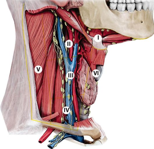 Рис. 557. Глубокие лимфатические узлы шеи, вид сбоку
Глубокие шейные лимфатические узлы.
Вид справа. Глубокие лимфатические узлы шеи состоят в основном из собирательных узлов. Они имеют важное клиническое значение как потенциальные пути метастазирования опухолей головы и шеи. Пораженные глубокие лимфатические узлы могут быть хирургически удалены или подвергнуты местному облучению. С клинической точки зрения глубокие шейные лимфатические узлы разделены на шесть групп:
I. Подподбородочные и поднижнечелюстные лимфатические узлы.
II - IV. Глубокие шейные лимфатические узлы, расположенные вдоль внутренней яремной вены (латеральные яремные лимфатические узлы).
II. Глубокие шейные лимфатические узлы (верхняя латеральная группа).
III. Глубокие шейные лимфатические узлы (средняя латеральная группа).
IV. Глубокие шейные лимфатические узлы (нижняя латеральная группа).
V. Лимфатические узлы заднего треугольника шеи.
VI. Передние шейные лимфатические узлы (передняя группа шейных узлов).
 Рис. 558. Отток лимфы от шеи, вид сбоку (схема):
1 - Axillary lymph nodes; 2 - Accessory nerve [XI]; Lymph nodes et vessels; 3 - Nuchal nodes; 4 - Occipital nodes; 5 - Deep parotid nodes; Pre-auricular nodes; 6 - Facial nodes; 7 - Submental nodes, submandibular nodes; 8 - Capillary of larynx, trachea, thyroid gland
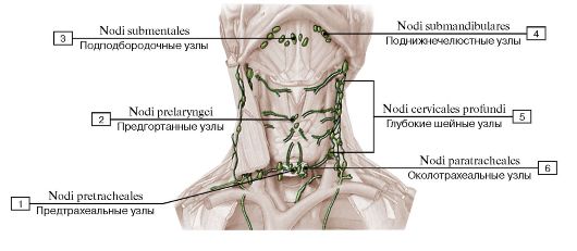 Рис. 559. Отток лимфы от шеи, вид спереди (схема):
1 - Pretracheal nodes; 2 - Prelaryngeal nodes; 3 - Submental nodes; 4 - Submandibular nodes; 5 - Deep cervical nodes; 6 - Paratracheal
nodes
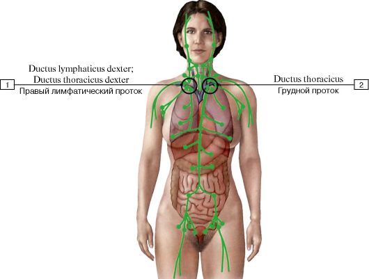 Рис. 560. Взаимоотношения лимфатических узлов шеи
с лимфатическими протоками, вид спереди:
1 - Right lymphatic duct; Right thoracic duct; 2 - Thoracic duct
Шейные лимфатические узлы могут быть затронуты заболеваниями, первично не воздействующими на область головы и шеи, так как лимфа оттекает от всего тела в правый и левый углы, образованные впадением внутренних яремных вен в подключичные вены. Это может привести к ретроградному вовлечению шейных узлов. Правый лимфатический проток оканчивается в правом углу, грудной проток - в левом. Кроме головных и шейных притоков, шейных узлов через грудной проток может достигнуть лимфа от грудных лимфатических узлов (средостенных и трахеобронхиальных), абдоминальных и каудальных лимфатических узлов. В результате заболевания этих органов возможно увеличение шейных лимфатических узлов.
Рак желудка может метастазировать в левую надключичную группу лимфатических узлов, образуя «предостерегающий узел», который подсказывает об абдоминальной опухоли. Системная лимфома может также распространиться на шейные лимфатические узлы этим путем.
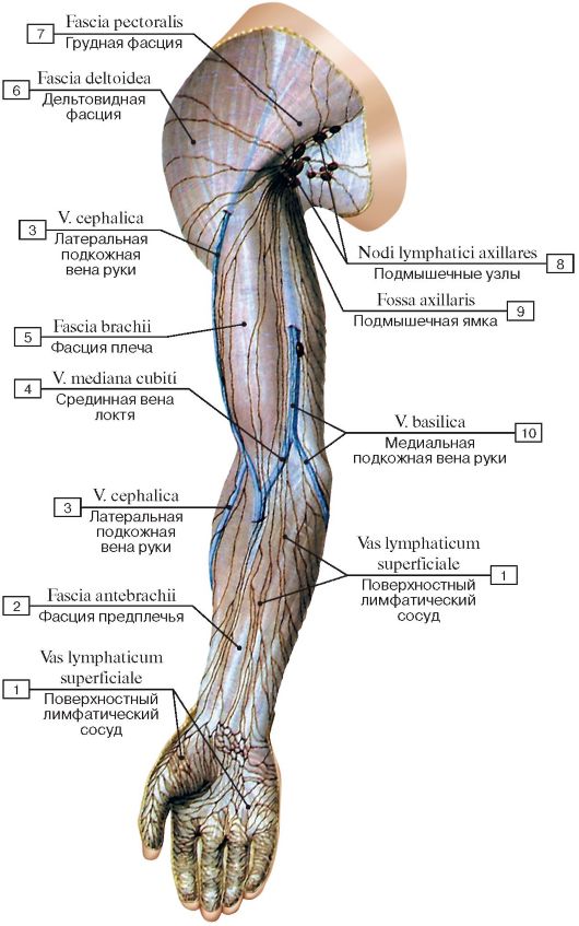 Рис. 561. Поверхностные лимфатические сосуды верхней конечности, вид спереди:
1 - Superficial lymph vessel; 2 - Antebrachial fascia; 3 - Cephalic vein; 4 - Median cubital vein; 5 - Brachial fascia; 6 - Deltoid fascia; 7 - Pectoral fascia; 8 - Axillary lymph nodes; 9 - Axillary fossa; 10 - Basilic vein
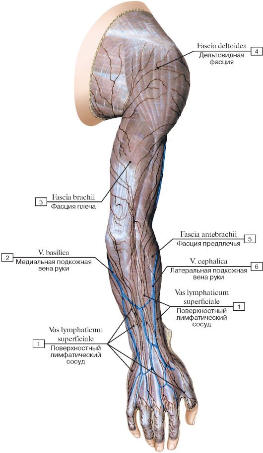 Рис. 562. Поверхностные лимфатические сосуды верхней конечности,
вид сзади:
1 - Superficial lymph vessel; 2 - Basilic vein; 3 - Brachial fascia; 4 - Deltoid fascia; 5 - Antebrachial fascia; 6 - Cephalic vein
Рис. 563. Глубокие лимфатические сосуды верхней конечности, вид спереди:
1 - Proper palmar digital arteries; 2 - Thenar eminence; 3 - Deep lymph vessel; 4 - Radial artery; 5 - Radial veins; 6 - Brachioradialis; 7 - Cubital nodes; 8 - Biceps brachii; 9 - Brachial artery; 10 - Cephalic vein; 11 - Pectoralis major; 12 - Axillary artery; Axillary vein; 13 - Subclavian artery; Subclavian vein; 14 - Scalenus anterior; Anterior scalene; 15 - Internal jugular vein; 16 - Brachiocephalic vein; 17 - Subclavian trunk; 18 - Pectoralis minor; 19 - Axillary lymph nodes; 20 - Brachial veins; 21 - Triceps brachii; 22 - Median cubital vein; 23 - Flexor carpi ulnaris; 24 - Ulnar veins; 25 - Anterior interosseous artery; Anterior interosseous veins; 26 - Ulnar artery; 27 - Deep palmar arch; 28 - Hypothenar eminence; 29 - Superficial palmar arch; 30 - Capillary of digits of hand
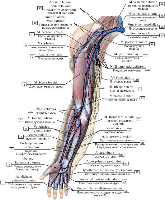
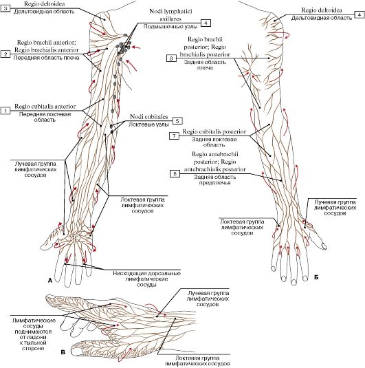 Рис. 564. Отток лимфы от верхней конечности (А - передняя поверхность, Б - задняя поверхность, В - кисть):
1 - Anterior region of elbow; 2 - Anterior region of arm; 3 - Deltoid region; 4 - Axillary lymph nodes; 5 - Cubital nodes; 6 - Posterior region of forearm; 7 - Posterior region of elbow; 8 - Posterior region of arm
Лимфатические сосуды верхней конечности подразделяются на два группы:
• Поверхностные лимфатические сосуды (vasa lymphatica superficialia)
• Глубокие лимфатические сосуды (vasa lymphatica profunda)
Глубокие лимфатические сосуды верхней конечности сопровождают артерии и глубокие вены, поверхностные залегают под кожей. На предплечье они наиболее тесно сопряжены с латеральной и медиальной подкожными венами руки. Между глубокой и поверхностной системами существуют многочисленные анастомозы. Стрелки на рисунке
показывают основные направления оттока лимфы. Воспалительные процессы руки вызывают опухание подмышечных лимфатических узлов и болезненность. Когда в воспалительные процессы вовлечены лимфатические сосуды, последние становятся заметными в виде красных полосок под кожей (лимфангит).
От большого и указательного пальцев и части среднего пальца лимфа оттекает по сосудам лучевой группы, которые достигают подмышечных лимфатических узлов. От остальных пальцев лимфа оттекает по локтевой группе лимфатических сосудов (здесь не показаны), которые оканчиваются в локтевых лимфатических узлах.
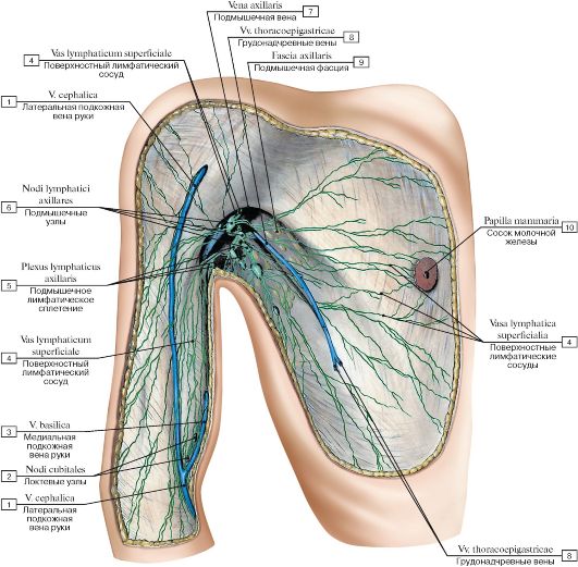 Рис. 565. Поверхностные лимфатические узлы и сосуды груди, плеча и подмышечной ямки:
1 - Cephalic vein; 2 - Cubital nodes; 3 - Basilic vein; 4 - Superficial lymph vessels; 5 - Axillary lymphatic plexus; 6 - Axillary lymph nodes;
7 - Axillary vein; 8 - Thoraco-epigastric veins; 9 - Axillary fascia; 10 - Nipple
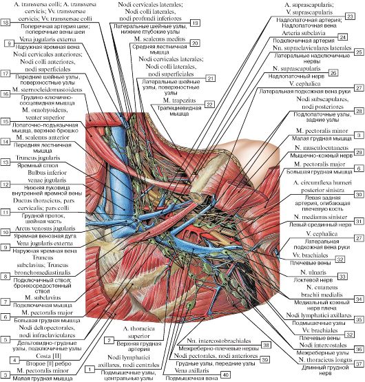 Рис. 566. Глубокие лимфатические узлы шеи и подмышечной ямки, левой:
1 - Axillary lymph nodes, central nodes; 2 - Superior thoracic artery; 3 - Pectoralis minor; 4 - Rib [II]; 5 - Deltopectoral nodes, infraclavicular nodes; 6 - Pectoralis major; 7 - Subclavius; 8 - Subclavian trunk; Bronchomediastinal trunk; 9 - External jugular vein; 10 - Jugular venous arch; 11 - Thoracic duct, cervical part; 12 - Inferior bulb of jugular vein; 13 - Jugular trunk; 14 - Scalenus anterior; Anterior scalene; 15 - Omohyoid, superior belly; 16 - Sternocleidomastoid; 17 - Anterior cervical nodes, superficial nodes; 18 - Transverse cervical artery; Transverse cervical veins; 19 - Lateral cervical nodes, inferior deep nodes; 20 - Scalenus medius; Middle scalene; 21 - Lateral cervical nodes, superficial nodes; 22 - Trapezius; 23 - Suprascapular artery; Suprascapular vein; 24 - Subclavian artery; 25 - Lateral supraclavicular nerves; 26 - Suprascapular nerve; 27 - Cephalic vein; 28 - Subscapular nodes, posterior nodes; 29 - Musculocutaneous nerve; 30 - Left posterior circumflex humeral artery; 31 - Left median nerve; 32 - Brachial veins; 33 - Ulnar nerve; 34 - Medial cutaneous nerve of arm; Medial brachial cutaneous nerve; 35 - Axillary lymph nodes; 36 - Intercostal nodes; 37 - Long thoracic nerve; 38 - Intercostobrachial
nerves; 39 - Pectoral nodes, anterior nodes; 40 - Axillary vein
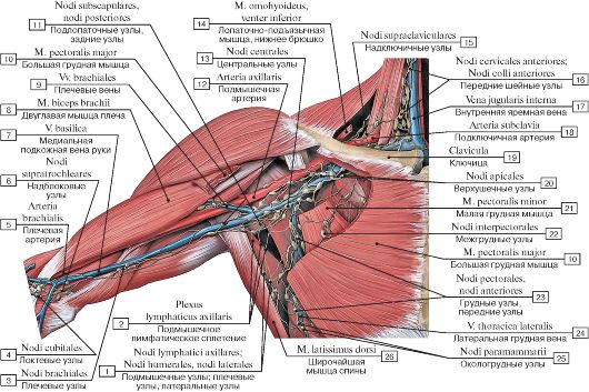 Рис. 567. Глубокие лимфатические узлы шеи и подмышечной ямки, правой:
1 - Axillary lymph nodes; Humeral nodes, lateral nodes; 2 - Axillary lymphatic plexus; 3 - Brachial nodes; 4 - Cubital nodes; 5 - Brachial artery; 6 - Supratrochlear nodes; 7 - Basilic vein; 8 - Biceps brachii; 9 - Brachial veins; 10 - Pectoralis major; 11 - Subscapular nodes, posterior nodes; 12 - Axillary artery; 13 - Central nodes; 14 - Omohyoid, inferior belly; 15 - Supraclavicular nodes; 16 - Anterior cervical nodes; 17 - Internal jugular vein; 18 - Subclavian artery; 19 - Clavicle; 20 - Apical nodes; 21 - Pectoralis minor; 22 - Interpectoral nodes; 23 - Pectoral nodes, anterior nodes; 24 - Lateral thoracic vein; 25 - Paramammary nodes; 26 - Latissimus dorsi
Подмышечные лимфатические узлы играют большую диагностическую роль при раке молочной железы. Злокачественная опухоль метастазирует в подмышечные узлы. В зависимости от характера метастазирования и показаний к их хирургическому удалению подмышечные лимфатические узлы подразделяют на три уровня в зависимости от их взаимоотношения с малой грудной мышцей.
• I уровень: все лимфатические узлы, расположенные латеральнее малой грудной мышцы.
• II уровень: все лимфатические узлы, расположенные на уровне малой грудной мышцы.
• III уровень: все лимфатические узлы, расположенные медиальнее малой грудной мышцы
 Рис. 568. Уровни расположения узлов подмышечной области:
1 - Pectoralis minor; 2 - Right lymphatic duct; Right thoracic duct
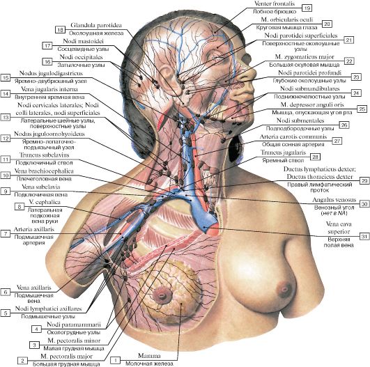 Рис. 569. Лимфатические узлы молочной железы, шеи и головы женщины:
1 - Breast; 2 - Pectoralis major; 3 - Pectoralis minor; 4 - Paramammary nodes; 5 - Axillary lymph nodes; 6 - Axillary vein; 7 - Axillary artery; 8 - Cephalic vein; 9 - Subclavian vein; 10 - Brachiocephalic vein; 11 - Subclavian trunk; 12 - Jugulo-omohyoid node; 13 - Lateral cervical nodes, superficial nodes; 14 - Internal jugular vein; 15 - Jugulo-digastric node; 16 - Occipital nodes; 17 - Mastoid nodes; 18 - Parotid gland; 19 - Frontal belly; 20 - Orbicularis oculi; 21 -Superficial parotid nodes; 22 - Zygomaticus major; 23 - Deep parotid nodes; 24 - Submandibular nodes; 25 - Depressor anguli oris; 26 - Submental nodes; 27 - Common carotid artery; 28 - Jugular trunk; 29 - Right lymphatic duct; Right thoracic duct; 30 - Venous angle; 31 - Superior vena cava
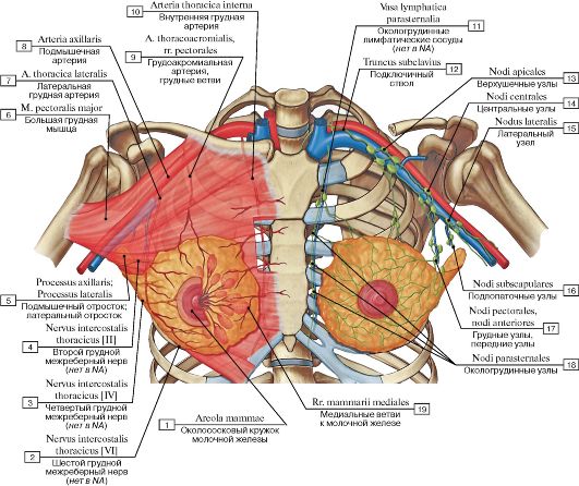 Рис. 570. Лимфатические сосуды и узлы молочной железы (схема):
1 - Areola; 2 - Thoracic intercostal nerve [VI]; 3 - Thoracic intercostal nerve[IV]; 4 - Thoracic intercostal nerve [II]; 5 - Axillary process; Axillary tail; 6 - Pectoralis major; 7 - Lateral thoracic artery; 8 - Axillary artery; 9 - Thoraco-acromial artery, pectoral branches; 10 - Internal thoracic artery; 11 - Parasternal lymphatic vessels; 12 - Subclavian trunk; 13 - Apical nodes; 14 - Central nodes; 15 - Lateral node; 16 - Subscapular nodes; 17 - Pectoral nodes, anterior nodes; 18 - Parasternal nodes; 19 - Medial mammary branches
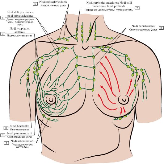 Рис. 571. Отток лимфы от молочной железы (указан стрелками):
1 - Submammary nodes; 2 - Paramammary nodes; 3 - Brachial nodes; 4 - Axillary lymph nodes; 5 - Deltopectoral nodes, infraclavicular nodes; 6 - Supraclavicular nodes; 7 - Anterior cervical nodes, deep nodes; 8 - Parasternal nodes
 Рис. 572. Лимфатические узлы тимуса:
1 - Brachiocephalic nodes; 2 - Right bronchomediastinal trunk; 3 - Left bronchomediastinal trunk; 4 - Right subclavian and internal jugular veins, joint; 5 - Left subclavian and internal jugular veins,
joint
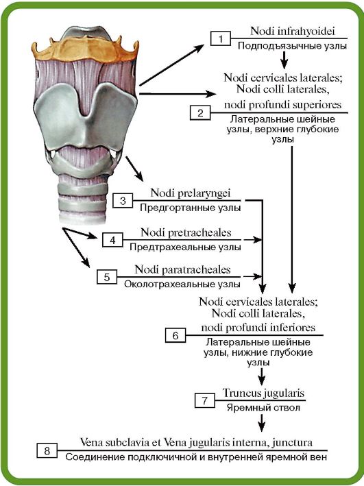 Рис. 573. Лимфатические узлы гортани:
1 - Infrahyoid nodes; 2 - Lateral cervical nodes, superior deep nodes; 3 - Prelaryngeal nodes; 4 - Pretracheal nodes; 5 - Paratracheal nodes; 6 - Lateral cervical nodes, inferior deep nodes; 7 - Jugular trunk;8 - Subclavian and Internal jugular veins, joint
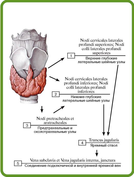 Рис. 574. Лимфатические узлы щитовидной железы:
1 - Lateral cervical superior deep nodes; 2 - Lateral cervical inferior deep nodes; 3 - Pretracheal and Paratracheal nodes; 4 - Jugular trunk; 5 - Subclavian and Internal jugular veins, joint
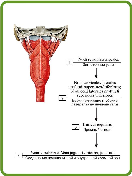 Рис. 575. Лимфатические узлы глотки:
1 - Retropharyngeal nodes; 2 - Lateral cervical superior/inferior deep nodes; 3 - Jugular trunk; 4 - Subclavian and Internal jugular veins, joint
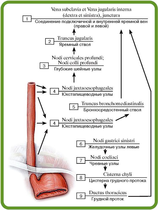 Рис. 576. Лимфатические узлы пищевода:
1 - Subclavian and Internal jugular veins (right and left), joint;
2 - Jugular trunk; 3 - Cervical deep nodes; 4 - Juxta-oesophageal nodes; 5 - Bronchomediastinal trunk; 6 - Left gastric nodes; 7 - Coeliac ganglia; 8 - Cisterna chyli; Chyle cistern; 9 - Thoracic
duct
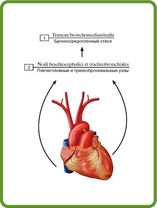 Рис. 577. Лимфатические узлы сердца:
1 - Bronchomediastinal trunk; 2 - Brachiocephalic and Tracheobronchial nodes
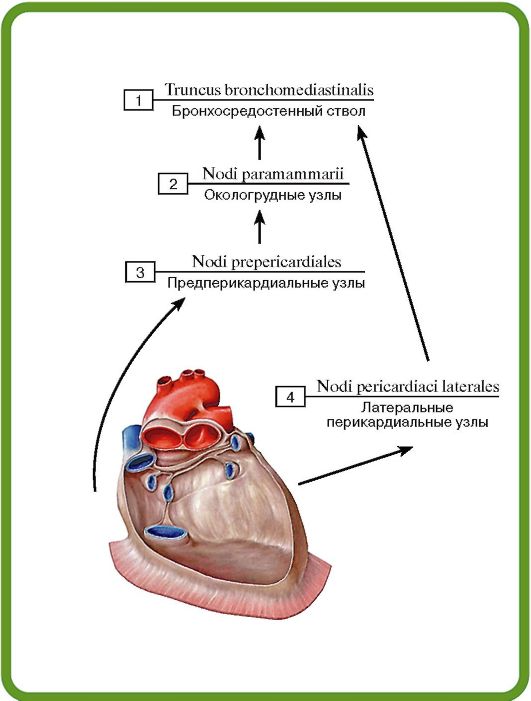 Рис. 578. Лимфатические узлы перикарда:
1 - Bronchomediastinal trunk; 2 - Paramammary nodes; 3 - Prepericardial nodes; 4 - Lateral pericardial nodes
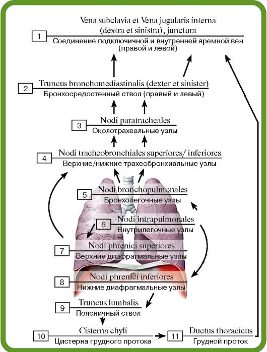 Рис. 579. Лимфатические узлы трахеи и легких:
1 - Subclavian and Internal jugular veins (right and left), joint;
2 - Bronchomediastinal trunk (right and left); 3 - Paratracheal nodes; 4 - Superior/inferior tracheobronchial nodes; 5 - Bronchopulmonary nodes; 6 - Intrapulmonary nodes; 7 - Superior diaphragmatic nodes; 8 - Inferior diaphragmatic nodes; 9 - Lumbar
trunk; 10 - Cisterna chyli; Chyle cistern; 11 - Thoracic duct
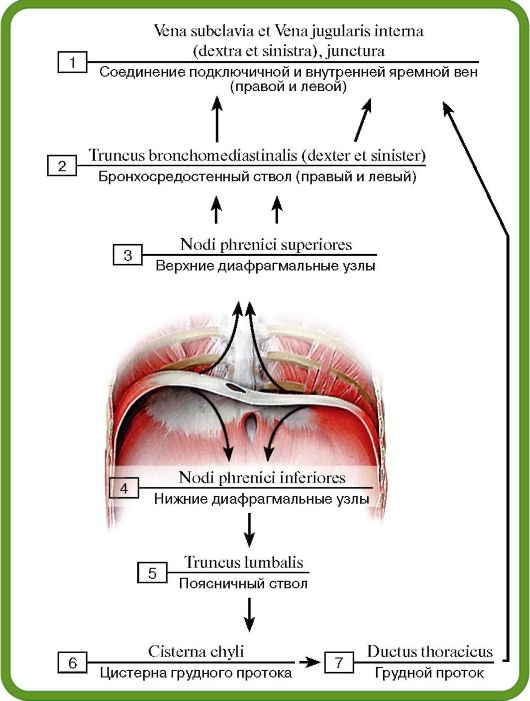 Рис. 580. Лимфатические узлы диафрагмы:
1 - Subclavian and Internal jugular veins (right and left), joint;
2 - Bronchomediastinal trunk (right and left); 3 - Superior diaphragmatic nodes; 4 - Inferior diaphragmatic nodes; 5 - Lumbar
trunk; 6 - Cisterna chyli; Chyle cistern; 7 - Thoracic duct
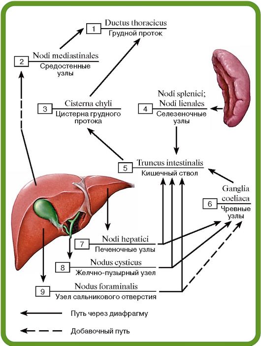 Рис. 581. Лимфатические узлы печени, желчного пузыря и селезенки:
1 - Thoracic duct; 2 - Mediastinal nodes; 3 - Cisterna chyli; Chyle cistern; 4 - Splenic nodes; 5 - Intestinal trunk; 6 - Coeliac nodes; 7 - Hepatic nodes; 8 - Cystic node; 9 - Node of anterior border of omental foramen
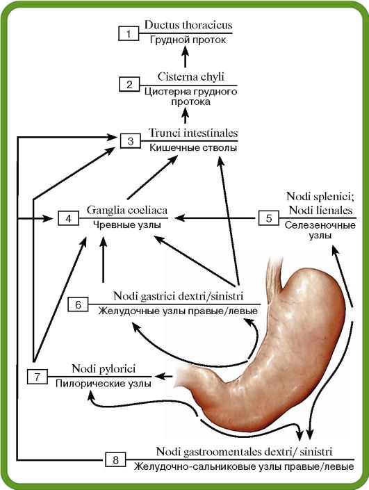 Рис. 582. Лимфатические узлы желудка:
1 - Thoracic duct; 2 - Cisterna chyli; Chyle cistern; 3 - Intestinal trunks; 4 - Coeliac nodes; 5 - Splenic nodes; 6 - Right/left gastric nodes; 7 - Pyloric nodes; 8 - Right/left gastro-omental nodes
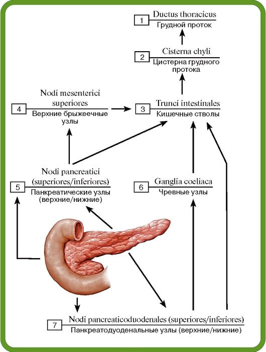 Рис. 583. Лимфатические узлы двенадцатиперстной кишки и поджелудочной железы:
1 - Thoracic duct; 2 - Cisterna chyli; Chyle cistern; 3 - Intestinal trunks; 4 - Superior mesenteric nodes; 5 - Pancreatic nodes (supe- rior/inferior); 6 - Coeliac nodes; 7 - Pancreaticoduodenal nodes (superior/inferior)
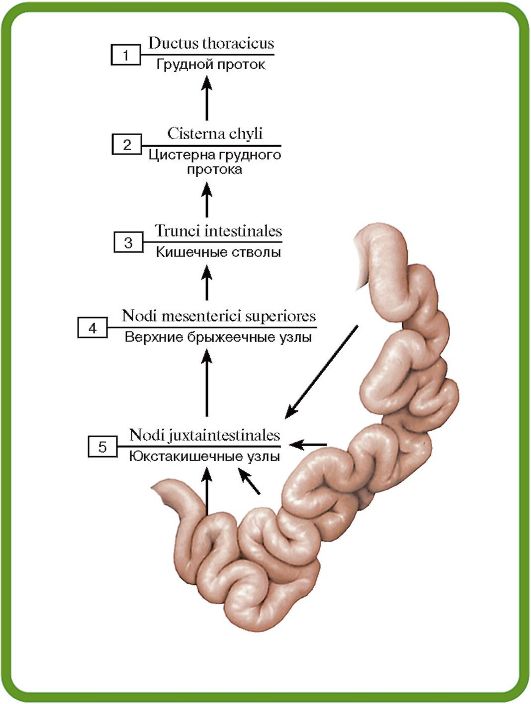 Рис. 584. Лимфатические узлы тощей и подвздошной кишок:
1 - Thoracic duct; 2 - Cisterna chyli; Chyle cistern; 3 - Intestinal trunks; 4 - Superior mesenteric nodes; 5 - Juxta-intestinal mesenteric nodes
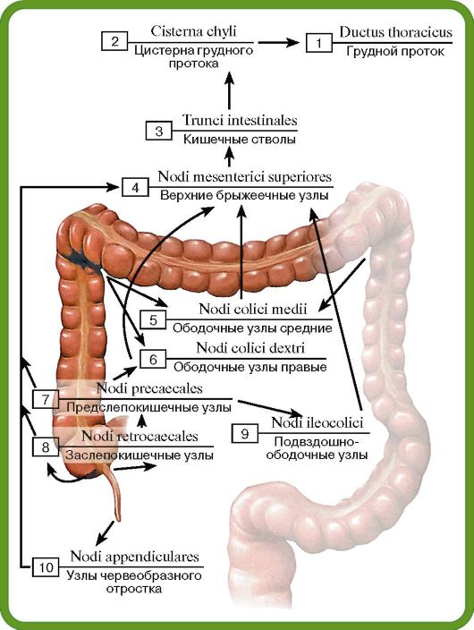 Рис. 585. Лимфатические узлы слепой кишки, аппендикса, восходящей и поперечной ободочн^1х кишок:
1 - Thoracic duct; 2 - Cisterna chyli; Chyle cistern; 3 - Intestinal trunks; 4 - Superior mesenteric nodes; 5 - Middle colic nodes; 6 - Right colic nodes; 7 - Precaecal nodes; 8 - Retrocaecal nodes; 9 - Ileocolic nodes; 10 - Appendicular nodes
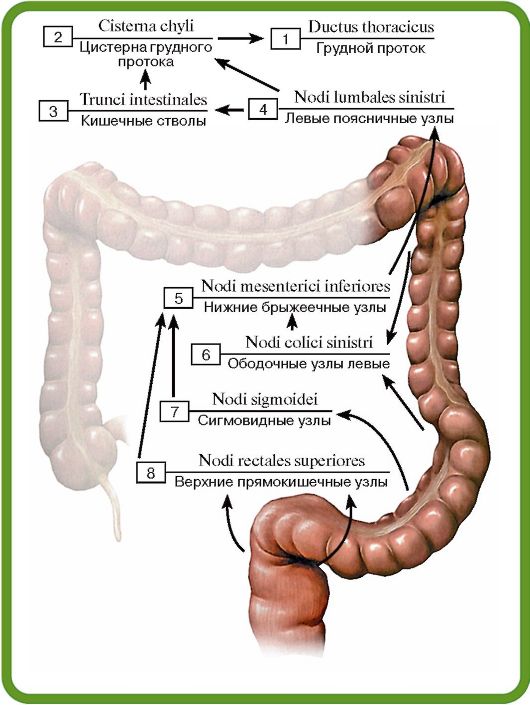 Рис. 586. Лимфатические узлы нисходящей и сигмовидной ободочных кишок:
1 - Thoracic duct; 2 - Cisterna chyli; Chyle cistern; 3 - Intestinal trunks; 4 - Left lumbar nodes; 5 - Inferior mesenteric nodes; 6 - Left colic nodes; 7 - Sigmoid nodes; 8 - Superior rectal nodes
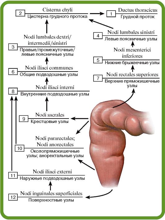 Рис. 587. Лимфатические узлы прямой кишки:
1 - Thoracic duct; 2 - Cisterna chyli; Chyle cistern; 3 - Right/inter- mediate/left lumbar nodes; 4 - Left lumbar nodes; 5 - Inferior mesenteric nodes; 6 - Common iliac nodes; 7 - Superior rectal nodes; 8 - Internal iliac nodes; 9 - Sacral nodes; 10 - Pararectal nodes: 11 - External iliac nodes; 12 - Superficial inguinal nodes
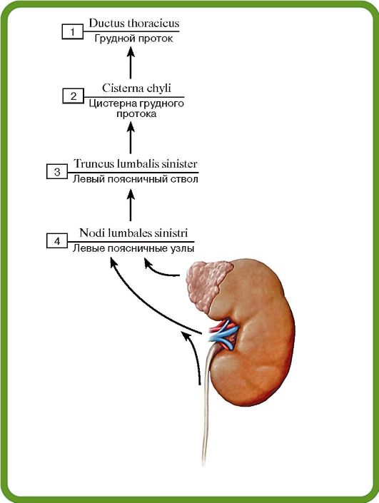 Рис. 588. Лимфатические узлы левой почки, мочеточника и надпочечника:
1 - Thoracic duct; 2 - Cisterna chyli; Chyle cistern; 3 - Left lumbar trunk; 4 - Left lumbar nodes
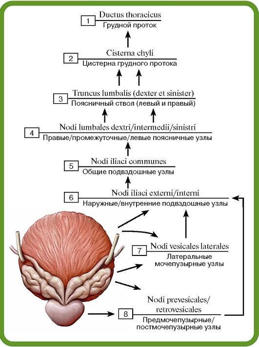 Рис. 589. Лимфатические узлы мочевого пузыря, простаты и семенных пузырьков:
1 - Thoracic duct; 2 - Cisterna chyli; Chyle cistern; 3 - Lumbar trunk (right and left); 4 - Right/intermediate/left lumbar nodes; 5 - Common iliac nodes; 6 - External/internal iliac nodes; 7 - Lateral vesical nodes; 8 - Prevesical/postvesical nodes
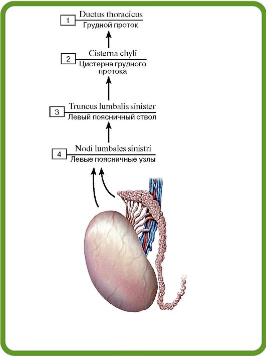 Рис. 590. Лимфатические узлы яичка, придатка яичка и семявыносящего протока:
1 - Thoracic duct; 2 - Cisterna chyli; Chyle cistern; 3 - Left lumbar trunk; 4 - Left lumbar nodes
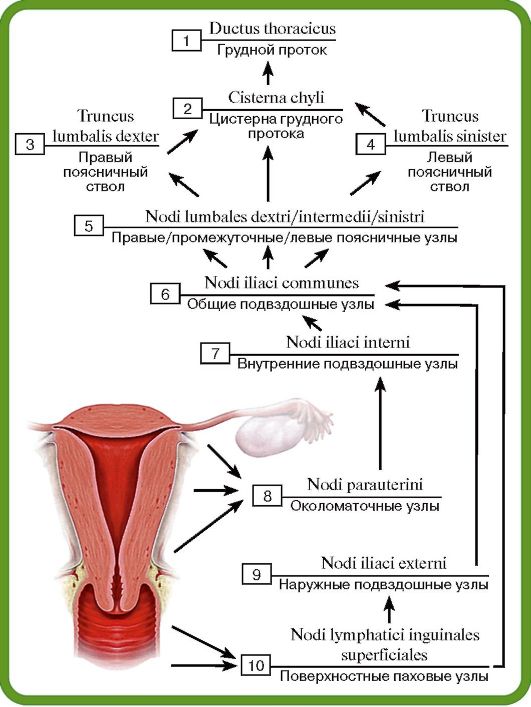 Рис. 591. Лимфатические узлы матки, маточной трубы и влагалища:
1 - Thoracic duct; 2 - Cisterna chyli; Chyle cistern; 3 - Right lumbar trunk; 4 - Left lumbar trunk; 5 - Right/intermediate/left lumbar nodes; 6 - Common iliac nodes; 7 - Internal iliac nodes; 8 - Parauterine nodes; 9 - External iliac nodes; 10 - Superficial inguinal
nodes
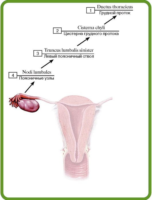 Рис. 592. Лимфатические узлы маточной трубы и яичника:
1 - Thoracic duct; 2 - Cisterna chyli; Chyle cistern; 3 - Left lumbar trunk; 4 - Lumbar nodes