Анатомия человека: атлас. Билич Г.Л., Крыжановский В.А. - Том 2. Внутренние органы. В 3-х томах.
|
|
|
|
СИСТЕМА СКЕЛЕТА
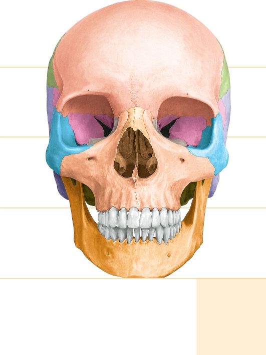
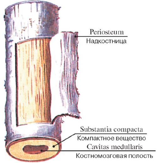 Рис. 1. Строение диафиза трубчатой кости
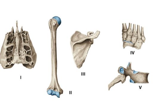 Рис. 2. Различные виды костей:
I - воздухоносная кость (решетчатая кость); II - длинная (трубчатая) кость; III - плоская кость; IV - губчатые (короткие) кости; V - смешанная кость
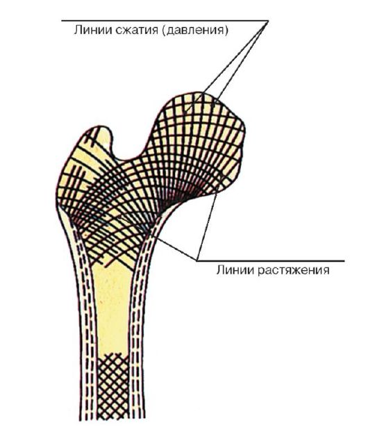 Рис. 3. Расположение костных перекладин в губчатом веществе (по линиям сжатия и растяжения)
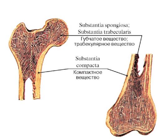 Рис. 4. Взаимоотношение компактного и губчатого веществ у проксимального и дистального эпифизов бедренной кости
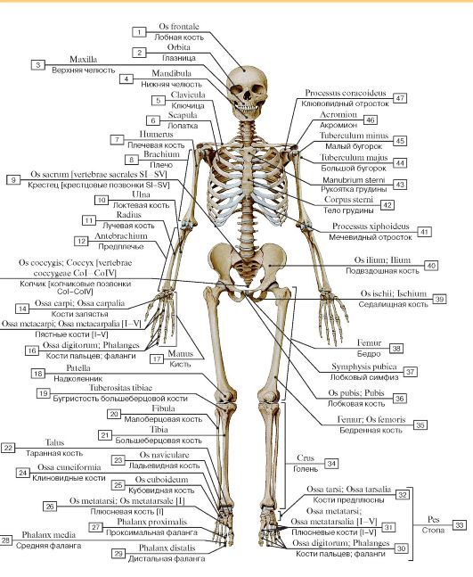 Рис. 5. Скелет человека, вид спереди:
1 - Frontal bone; 2 - Orbit; 3 - Maxilla; 4 - Mandible; 5 - Clavicle; 6 - Scapula; 7 - Humerus; 8 - Arm; 9 - Sacrum [sacral vertebrae SI-SV]; 10 - Ulna; 11 - Radius; 12 - Forearm; 13 - Coccyx [coccygeal vertebrae CoI-CoIV]; 14 - Carpal bones; 15 - Metacarpals [I-V]; 16 - Phalanges; 17 - Hand; 18 - Patella; 19 - Tibial tuberosity; 20 - Fibula; 21 - Tibia; 22 - Talus; 23 - Navicular; 24 - Cuneiform bone; 25 - Cuboid; 26 - Metatarsal [I]; 27 - Proximal phalanx; 28 - Middle phalanx; 29 - Distal phalanx; 30 - Phalanges; 31 - Metatarsals [I-V]; 32 - Tarsal bones; 33 = 30 + 31 + 32 - Foot; 34 - Leg; 35 - Femur; Thigh bone; 36 - Pubis; 37 - Pubic symphysis; 38 - Thigh; 39 - Ischium; 40 - Ilium; 41 - Xiphoid process; 42 - Body of sternum; 43 - Manubrium of sternum; 44 - Greater tubercle; 45 - Lesser tubercle;
46 - Acromion; 47 - Coracoid process
 Рис. 6. Скелет человека, вид сзади:
1 - Atlas [CI]; 2 - Axis [CII]; 3 - Scapula; 4 - Spine of scapula; 5 - Acromion; 6 - Humerus; 7 - Iliac crest; 8 - Olecranon; 9 - Head of radius; 10 - Acetabulum; 11 - Ulna; 12 - Radius; 13 - Ulnar styloid process; 14 - Pisiform; 15 - Head of femur; 16 - Sacrum [sacral vertebrae SI-SV]; 17 - Linea aspera; 18 - Medial condyle; 19 - Lateral condyle; 20 - Head of fibula; 21 - Head of tibia; 22 - Medial malleolus; 23 - Lateral malleolus; 24 - Talus; 25 - Calcaneus; 26 - Tibia; 27 - Fibula; 28 - Lesser trochanter; 29 - Neck of femur; 30 - Greater trochanter; 31 - Hamate; 32 - Triquetrum; 33 - Capitate; 34 - Trapezoid; 35 - Trapezium; 36 - Scaphoid; 37 - Lunate; 38 - Vertebral column; 39 - Head of humerus; 40 - Occipital bone; 41 - Parietal bone
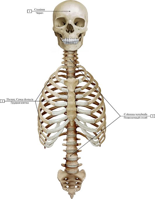 Рис. 7. Осевой скелет, вид спереди:
1 - Cranium; 2 - Thorax; Thoracic cage; 3 - Vertebral column
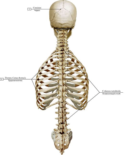 Рис. 8. Осевой скелет, вид сзади:
1 - Cranium; 2 - Thorax; Thoracic cage; 3 - Vertebral column
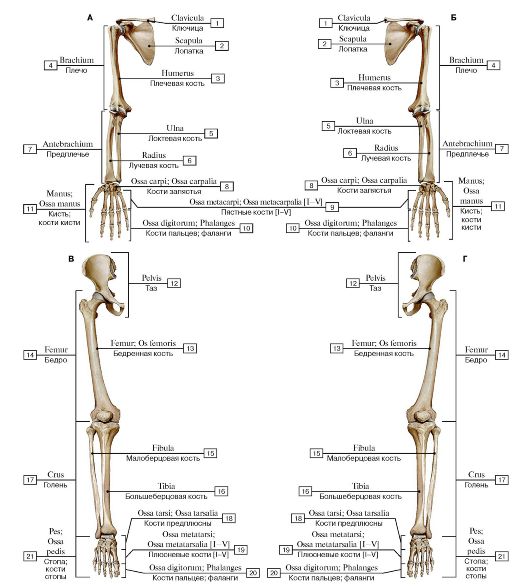 Рис.
9. Добавочный скелет, вид спереди (А - правая верхняя конечность, Б -
левая верхняя конечность, В - правая нижняя конечность, Г - левая нижняя
конечность):
1 - Clavicle; 2 - Scapula; 3 - Humerus; 4 - Arm; 5 - Ulna; 6 - Radius; 7 - Forearm; 8 - Carpal bones; 9 - Metacarpals [I-V]; 10 - Phalanges; 11 - Hand; Bones of hand; 12 - Pelvis; 13 - Femur; Thigh bone; 14 - Thigh; 15 - Fibula; 16 - Tibia; 17 - Leg; 18 - Tarsal bones; 19 - Metatarsals [I-V]; 20 - Phalanges; 21 - Foot; Bones of foot
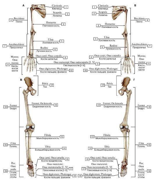 Рис.
10. Добавочный скелет, вид сзади (А - правая верхняя конечность, Б -
левая верхняя конечность, В - правая нижняя конечность, Г - левая нижняя
конечность):
1 - Clavicle; 2 - Scapula; 3 - Humerus; 4 - Arm; 5 - Ulna; 6 - Radius; 7 - Forearm; 8 - Carpal bones; 9 - Metacarpals [I-V]; 10 - Phalanges; 11 - Hand; Bones of hand; 12 - Pelvis; 13 - Femur; Thigh bone; 14 - Thigh; 15 - Fibula; 16 - Tibia; 17 - Leg; 18 - Tarsal bones; 19 - Metatarsals [I-V]; 20 - Phalanges; 21 - Foot; Bones of foot
 Рис. 11. Позвоночный столб (А - вид спереди, Б - вид сзади, В - вид сбоку, слева):
1 - Anterior sacral foramina; 2 - Coccyx [coccygeal vertebrae CoI-CoIV]; 3 - Sacrum [sacral vertebrae SI-SV]; 4 - Posterior sacral foramina; 5 - Lumbar vertebrae [LI-LV]; 6 - Transverse process; 7 - Thoracic vertebrae [TI-TXII]; 8 - Spinous process; 9 - Cervical vertebrae [CI-CVII]; 10 - Atlas [CI]; 11 - Axis [CII]; 12 - Vertebra prominens [CVII]; 13 - Superior costal facet; 14 - Inferior costal facet; 15 - Transverse costal facet; 16 - Superior articular process; 17 - Inferior articular process; 18 - Intervertebral foramen; 19 - Intervertebral disc;
20 - Promontory; 21 - Auricular surface
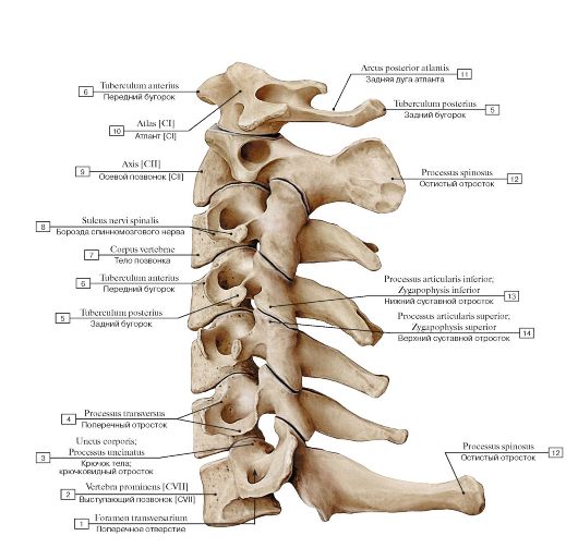 Рис. 12. Шейный отдел позвоночного столба, вид сбоку, слева:
1 - Foramen transversarium; 2 - Vertebra prominens [CVII]; 3 - Uncus of body; Uncinate process; 4 - Transverse process; 5 - Posterior tubercle; 6 - Anterior tubercle; 7 - Vertebral body; 8 - Groove for spinal nerve; 9 - Axis [CII]; 10 - Atlas [CI]; 11 - Posterior arch; 12 - Spinous process; 13 - Inferior articular process; 14 - Superior articular process
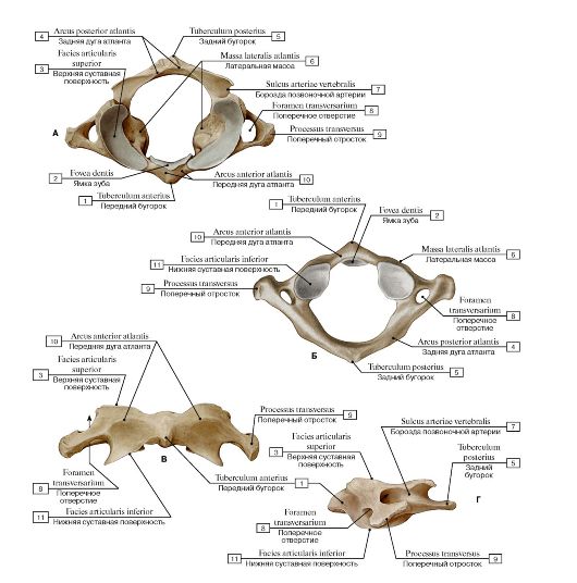 Рис. 13. Первый шейный позвонок, атлант [CI] (А - вид сверху, Б - вид снизу, В - вид спереди, Г - вид сбоку, слева):
1 - Anterior tubercle; 2 - Facet for dens; 3 - Superior articular surface; 4 - Posterior arch; 5 - Posterior tubercle; 6 - Lateral mass; 7 - Groove for vertebral artery; 8 - Foramen transversarium; 9 - Transverse process; 10 - Anterior arch; 11 - Inferior articular surface
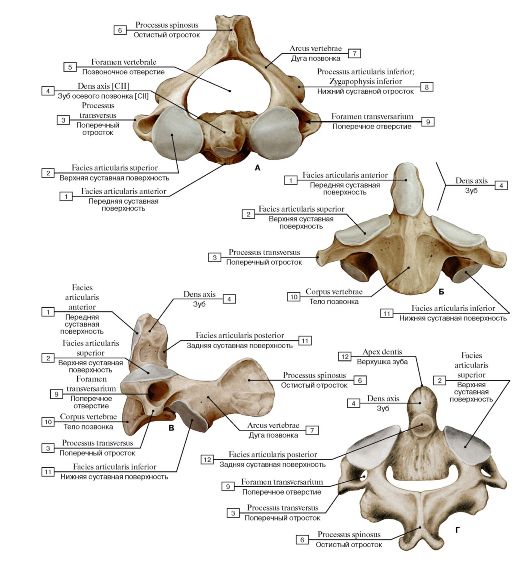 Рис. 14. Второй шейный позвонок, осевой [CII] (А - вид сверху, Б - вид спереди, В - вид сбоку, слева, Г - вид сзади):
1 - Anterior articular facet; 2 - Superior articular surface; 3 - Transverse process; 4 - Dens axis [CII]; 5 - Vertebral foramen; 6 - Spinous process; 7 - Vertebral arch; 8 - Inferior articular process; 9 - Foramen transversarium; 10 - Vertebral body; 11 - Posterior articular facet;
12 - Apex (Dens)
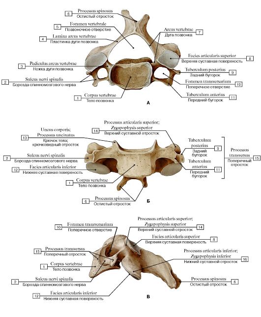 Рис. 15. Четвертый шейный позвонок [CIV] (А - вид сверху, Б - вид спереди, В - вид сбоку, слева):
1 - Vertebral body; 2 - Groove for spinal nerve; 3 - Pedicle; 4 - Lamina; 5 - Vertebral foramen; 6 - Spinous process; 7 - Vertebral arch; 8 - Superior articular facet; 9 - Posterior tubercle; 10 - Foramen transversarium; 11 - Anterior tubercle; 12 - Inferior articular facet; 13 - Uncus of body; Uncinate process; 14 - Superior articular process; 15 - Transverse process; 16 - Inferior articular process
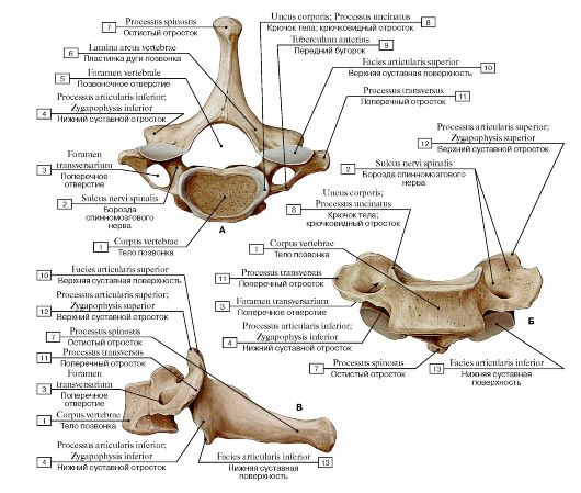 Рис. 16. Седьмой шейный позвонок [CVII] (А - вид сверху, Б - вид спереди, В - вид сбоку, слева):
1 - Vertebral body; 2 - Groove for spinal nerve; 3 - Foramen transversarium; 4 - Inferior articular process; 5 - Vertebral foramen; 6 - Lamina; 7 - Spinous process; 8 - Uncus of body; Uncinate process; 9 - Anterior tubercle; 10 - Superior articular facet; 11 - Transverse process; 12 - Superior articular process; 13 - Inferior articular facet
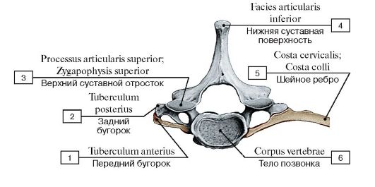 Рис. 17. Шейное ребро:
1 - Anterior tubercle; 2 - Posterior tubercle; 3 - Superior articular process; 4 - Inferior articular facet; 5 - Cervical rib; 6 - Vertebral body
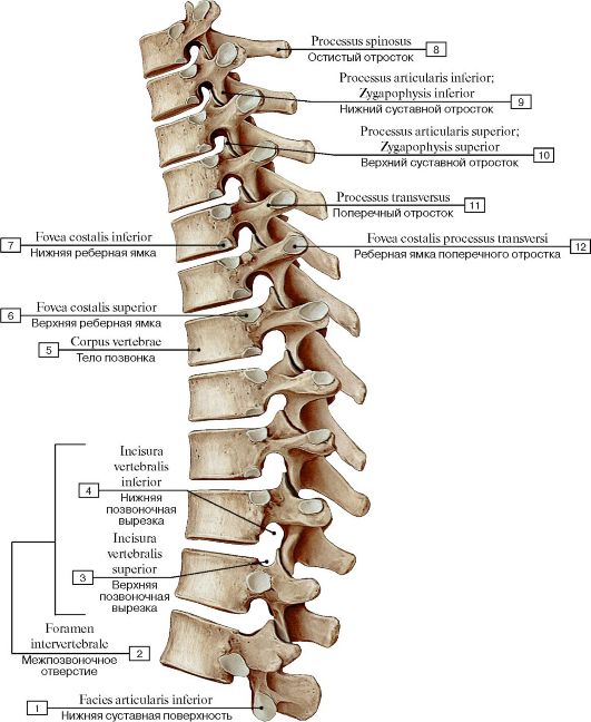 Рис. 18. Грудной отдел позвоночного столба, вид сбоку, слева:
1 - Inferior articular facet; 2 = 3 +4 - Intervertebral foramen; 3 - Superior vertebral notch; 4 - Inferior vertebral notch; 5 - Vertebral body; 6 - Superior costal fovea; 7 - Inferior costal fovea; 8 - Spinous process; 9 - Inferior articular process; 10 - Superior articular process;
11 - Transverse process; 12 - Transverse costal fovea
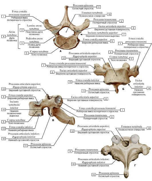 Рис. 19. Первый грудной позвонок [TI] (А - вид сверху, Б - вид спереди, В - вид сбоку, слева,
Г - вид сзади):
1 - Transverse costal facet; 2 - Lamina; 3 - Pedicle; 4 = 2 + 3 - Vertebral arch; 5 - Vertebral body; 6 - Superior costal facet; 7 - Superior vertebral notch; 8 - Superior articular facet; 9 - Transverse process; 10 - Vertebral foramen; 11 - Spinous process; 12 - Superior articular process; 13 - Inferior costal facet; 14 - Inferior articular facet; 15 - Inferior vertebral notch; 16 - Inferior articular process
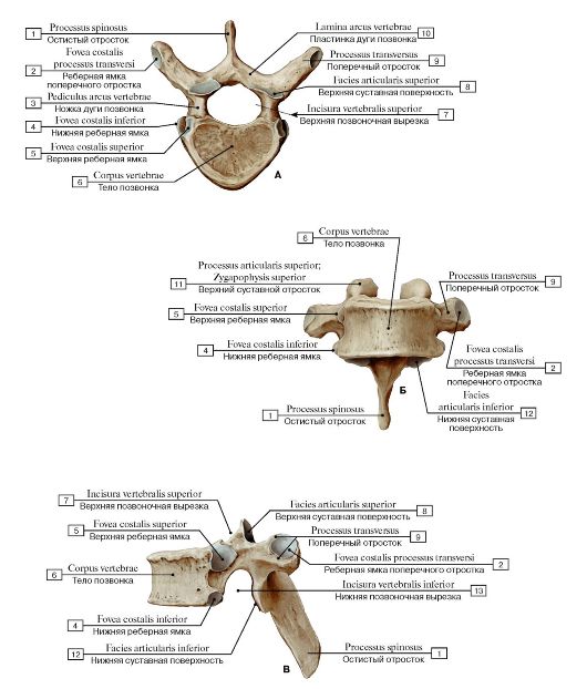 Рис. 20. Четвертый грудной позвонок [TIV] (А - вид сверху, Б - вид спереди, В - вид сбоку, слева):
1 - Spinous process; 2 - Transverse costal facet; 3 - Pedicle; 4 - Inferior costal facet; 5 - Superior costal facet; 6 - Vertebral body; 7 - Superior vertebral notch; 8 - Superior articular facet; 9 - Transverse process; 10 - Lamina; 11 - Superior articular process; 12 - Inferior articular
facet; 13 - Inferior vertebral notch
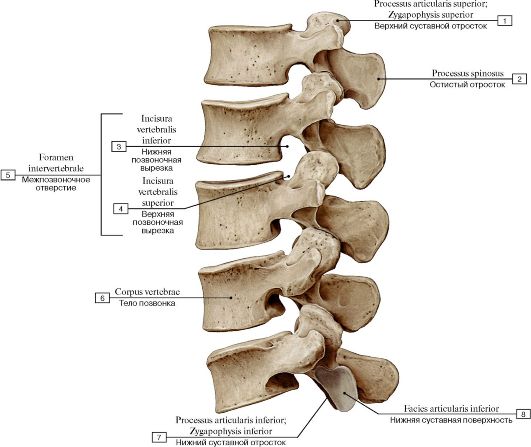 Рис. 21. Поясничный отдел позвоночного столба, вид сбоку, слева:
1 - Superior articular process; 2 - Spinous process; 3 - Inferior vertebral notch; 4 - Superior vertebral notch; 5 = 3 + 4 - Intervertebral foramen; 6 - Vertebral body; 7 - Inferior articular process; 8 - Inferior articular facet
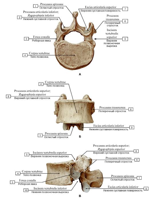 Рис. 22. Первый поясничный позвонок [LI] (А - вид сверху, Б - вид спереди, В - вид сбоку, слева):
1 - Spinous process; 2 - Inferior articular process; 3 - Costal facet; 4 - Vertebral body; 5- Superior vertebral notch; 6 - Transverse process; 7 - Superior articular facet; 8 - Superior articular process; 9 - Inferior articular facet; 10 - Inferior vertebral notch
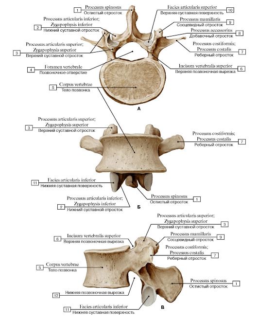 Рис. 23. Третий поясничный позвонок [LIII] (А - вид сверху, Б - вид спереди, В - вид сбоку, слева):
1 - Spinous process; 2 - Inferior articular process; 3 - Superior articular process; 4 - Vertebral foramen; 5 - Vertebral body; 6 - Superior vertebral notch; 7 - Costal process; 8 - Accessory process; 9 - Mammillary process; 10 - Superior articular facet; 11 - Inferior articular
facet; 12 - Inferior vertebral notch
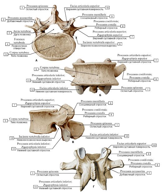 Рис. 24. Четвертый поясничный позвонок [LIV] (А - вид сверху, Б - вид спереди, В - вид сбоку, слева,
Г - вид сзади):
1 - Spinous process; 2 - Accessory process; 3 - Vertebral arch; 4 - Vertebral foramen; 5 - Vertebral body; 6 - Superior vertebral notch; 7 - Superior articular process; 8 - Costal process; 9 - Mammillary process; 10 - Superior articular surface; 11 - Inferior articular process; 12 - Inferior articular facet; 13 - Inferior vertebral notch
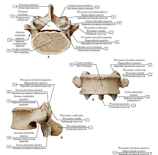 Рис. 25. Пятый поясничный позвонок [LV] (А - вид сверху, Б - вид спереди, В - вид сбоку, слева):
1 - Spinous process; 2 - Vertebral foramen; 3 - Superior vertebral notch; 4 - Vertebral body; 5 - Pedicle; 6 - Superior articular process; 7 - Costal process; 8 - Superior articular facet; 9 - Inferior articular process; 10 - Lamina; 11 - Inferior articular facet; 12 - Inferior vertebral notch
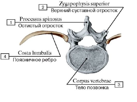 Рис. 26. Поясничное ребро:
1 - Spinous process; 2 - Superior articular process; 3 - Vertebral body; 4 - Lumbar rib
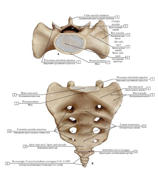 Рис. 27. Крестец и копчик (А - вид сверху, Б - вид спереди):
1 - Superior articular process; 2 - Median sacral crest; 3 - Sacral canal; 4 - Lateral part; 5 - Ala; Wing; 6 - Base of sacrum; 7 - Promontory; 8 - Coccyx [coccygeal vertebrae CoI-CoIV]; 9 - Apex; 10 - Anterior sacral foramina; 11 - Transverse ridges; 12 - Sacrococcygeal joint
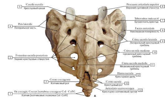
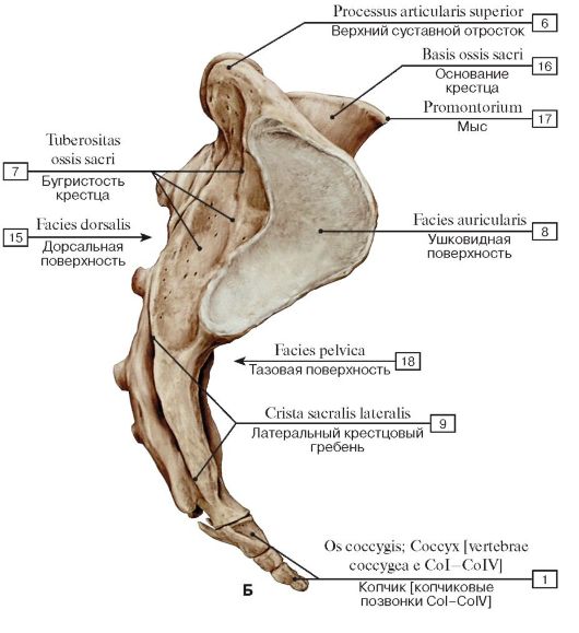 Рис. 28. Крестец и копчик (А - вид сзади, Б - вид сбоку, справа):
1 - Coccyx [coccygeal vertebrae CoI-CoIV]; 2 - Coccygeal cornu; 3 - Posterior sacral foramina; 4 - Lateral part; 5 - Sacral canal; 6 - Superior articular process; 7 - Sacral tuberosity; 8 - Auricular surface; 9 - Lateral sacral crest; 10 - Median sacral crest; 11 - Intermediate sacral crest; 12 - Sacral hiatus; 13 - Sacral cornu; Sacral horn; 14 - Sacrococcygeal joint; 15 - Dorsal surface; 16 - Base of sacrum;17 - Promontory; 18 - Pelvic surface
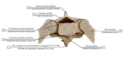 Рис. 29. Поперечное сечение на уровне вторых крестцовых отверстий:
1 - Coccyx [coccygeal vertebrae CoI-CoIV]; 2 - Anterior sacral foramina; 3 - Posterior sacral foramina; 4 - Sacral canal; 5 - Median sacral
crest; 6 - Lateral part; 7 - Pelvic surface
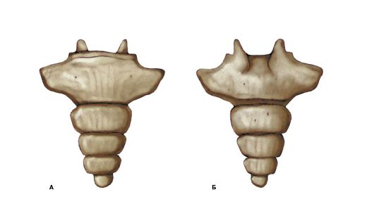 Рис. 30. Копчик [копчиковые позвонки CoI-CoIV] (А - вид спереди, Б - вид сзади)
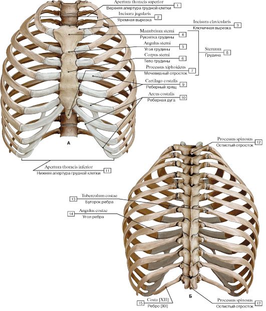 Рис. 31. Скелет грудной клетки (А - вид спереди, Б - вид сзади):
1 - Superior thoracic aperture; Thoracic inlet; 2 - Jugular notch; Suprasternal notch; 3 - Clavicular notch; 4 - Manubrium of sternum; 5 - Sternal angle; 6 - Body of sternum; 7 - Xiphoid process; 8 - Sternum; 9 - Costal cartilage; 10 - Costal margin; Costal arch; 11 - Inferior thoracic aperture; Thoracic outlet; 12 - Spinous process; 13 - Tubercle; 14 - Angle of rib; 15 - Rib [XII]
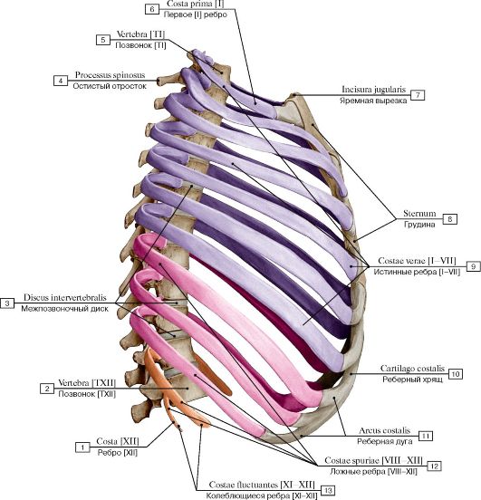 Рис. 32. Скелет грудной клетки, вид сбоку, справа:
1 - Rib [XII]; 2 - Vertebra [TXII]; 3 - Intervertebral disc; 4 - Spinous process; 5 - Vertebra [TI]; 6 - First rib [I]; 7 - Jugular notch; Suprasternal notch; 8 - Sternum; 9 - True ribs [I-VII]; 10 - Costal cartilage; 11 - Costal margin; Costal arch; 12 - False ribs [VIII-XII];
13 - Floating ribs [Xl-XII]
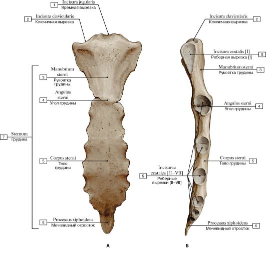 Рис. 33. Грудина (А - вид спереди, Б - вид сбоку, справа):
1 - Jugular notch; Suprasternal notch; 2 - Clavicular notch; 3 - Manubrium of sternum; 4 - Sternal angle; 5 - Body of sternum; 6 - Xiphoid process; 7 - Sternum; 8 - Constal notch [I]; 9 - Costal notches [II-VII]
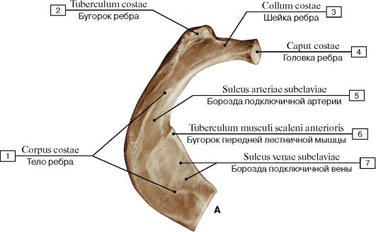
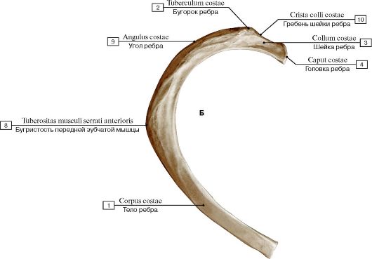 Рис. 34. Ребра (А - первое [I] ребро, правое, вид сверху; Б - второе [II] ребро, правое, вид сверху):
1 - Body of rib; Shaft of rib; 2 - Tubercle; 3 - Neck of rib; 4 - Head of rib; 5 - Groove for subclavian artery; 6 - Scalene tubercle; 7 - Groove for subclavian vein; 8 - Tuberosity for serratus anterior; 9 - Angle of rib; 10 - Crest of neck rib
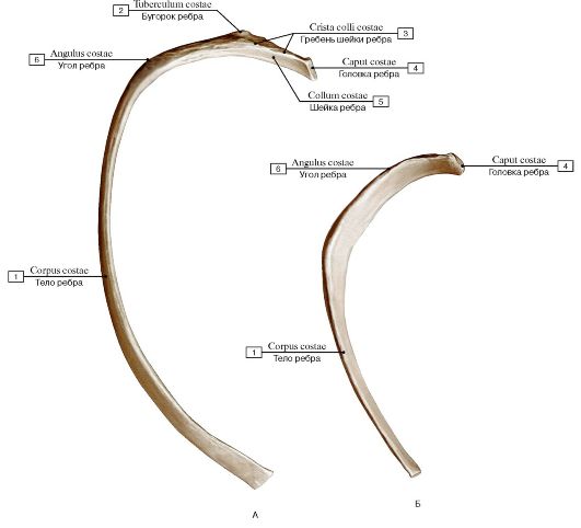 Рис. 35. Ребра (А - пятое ребро, правое; Б - одиннадцатое ребро, правое):
1 - Body of rib; Shaft of rib; 2 - Tubercle; 3 - Crest of neck rib; 4 - Head of rib; 5 - Neck of rib; 6 - Angle of rib
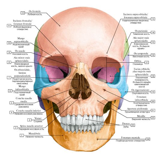 Рис. 36. Череп, вид спереди:
I - Mandible; 2 - Anterior nasal spine; 3 - Vomer; 4 - Inferior nasal concha; 5 - Middle nasal concha; 6 - Infra-orbital margin; 7 - Ethmoid; Ethmoidal bone, perpendicular plate; 8 - Sphenoid; Sphenoidal bone, lesser wing; 9 - Nasal bone; 10 - Supra-orbital margin;
II - Frontal notch/foramen; 12 - Frontal bone; 13 - Supra-orbital notch/foramen; 14 - Parietal bone; 15 - Sphenoid; Sphenoidal bone, greater wing; 16 - Temporal bone; 17 - Orbit; 18 - Orbital surface; Sphenoid; Sphenoidal bone, greater wing; 19 - Zygomatic bone;
20 - Infra-orbital foramen; 21 - Piriform aperture; 22 - Maxilla; 23 - Teeth; 24 - Mental foramen
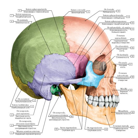 Рис. 37. Череп, вид сбоку, справа:
1 - External acoustic meatus; 2 - Temporal bone, mastoid process; 3 - Temporal bone, squamous part; 4 - Lambdoid suture; 5 - Occipital bone; 6 - Parietal bone; 7 - Squamous suture; 8 - Sphenoparietal suture; 9 - Coronal suture; 10 - Frontal bone; 11 - Sphenofrontal suture; 12 - Sphenosquamous suture; 13 - Sphenoid; Sphenoidal bone, greater wing; 14 - Supra-orbital notch/foramen; 15 - Ethmoid; Ethmoidal bone; 16 - Lacrimal bone; 17 - Nasal bone; 18 - Infra-orbital foramen; 19 - Maxilla; 20 - Mandible; 21 - Mental foramen; 22 - Zygomatic bone; 23 - Zygomatic arch; 24 - Temporal bone, styloid process
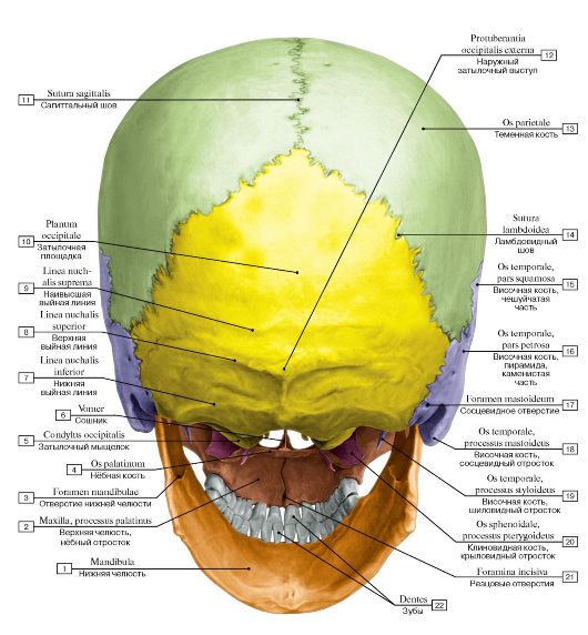 Рис. 38. Череп, вид сзади:
1 - Mandible; 2 - Maxilla, palatine process; 3 - Mandibular foramen; 4 - Palatine bone; 5 - Occipital condyle; 6 - Vomer; 7 - Inferior nuchal line; 8 - Superior nuchal line; 9 - Highest nuchal line; 10 - Occipital plane; 11 - Sagittal suture; 12 - External occipital protuberance; 13 - Parietal bone; 14 - Lambdoid suture; 15 - Temporal bone, squamous part; 16 - Temporal bone, petrous part; 17 - Mastoid foramen; 18 - Temporal bone, mastoid process; 19 - Temporal bone, styloid process; 20 - Sphenoid; Sphenoidal bone, pterygoid process; 21 - Incisive foramina; 22 - Teeth
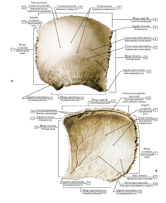 Рис. 39. Теменная кость, правая (А - вид снаружи, Б - вид изнутри):
I - Occipital border; 2 - Occipital angle;
3 - Parietal tuber; Parietal eminence;
4 - Parietal foramen; 5 - External surface; 6 - Sagittal border; 7 - Frontal angle; 8 - Superior temporal line; 9 - Inferior temporal line; 10 - Frontal border;
II - Sphenoidal angle; 12 - Squamosal border; 13 - Mastoid angle; 14 - Granular foveolae; 15 - Groove for superior sagittal sinus; 16 - Internal surface; 17 - Grooves for arteries; 18 - Groove for sigmoid
sinus
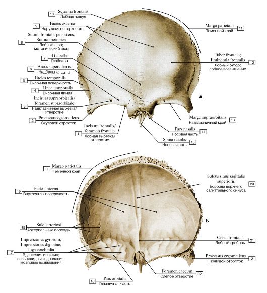 Рис. 40. Лобная кость (А - вид снаружи, Б - вид изнутри):
1 - Frontal notch/foramen; 2 - Zygomatic process; 3 - Supra-orbital notch/foramen; 4 - Temporal line; 5 - Temporal surface; 6 - Superciliary arch; 7 - Glabella; 8 - Frontal suture; Metopic suture; 9 - External surface; 10 - Squamous part; 11 - Parietal margin; 12 - Frontal tuber; Frontal eminence; 13 - Supra-orbital margin; 14 - Nasal part; 15 - Nasal spine; 16 - Orbital part; 17 - Impressions of cerebral gyri; 18 - Grooves for arteries; 19 - Internal surface; 20 - Groove for superior sagittal sinus; 21 - Frontal crest; 22 - Foramen caecum
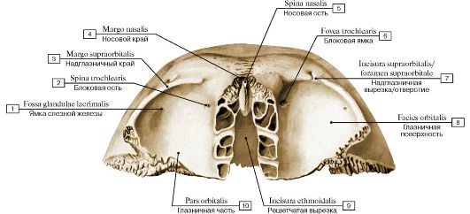 Рис. 41. Лобная кость, вид снизу
1 - Fossa for lacrimal gland; Lacrimal fossa; 2 - Trochlear spine; 3 - Supra-orbital margin; 4 - Nasal margin; 5 - Nasal spine; 6 - Trochlear fovea; 7 - Supra-orbital notch/foramen; 8 - Orbital surface; 9 - Ethmoidal notch; 10 - Orbital part
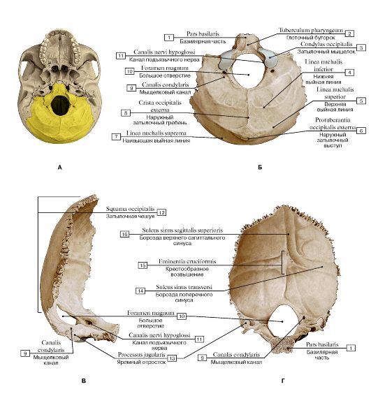 Рис.
42. Затылочная кость (А - затылочная кость в составе черепа - выделена
цветом, Б - вид снизу и сзади, В - вид сбоку, справа, Г - вид изнутри,
спереди):
1 - Basilar part; 2 - Pharyngeal tubercle; 3 - Occipital condyle; 4 - Inferior nuchal line; 5 - Superior nuchal line; 6 - External occipital protuberance; 7 - Highest nuchal line; 8 - External occipital crest; 9 - Condylar canal; 10 - Foramen magnum; 11 - Hypoglossal canal; 12 - Squamous part of occipital bone; 13 - Jugular process; 14 - Groove for transverse sinus; 15 - Cruciform eminence; 16 - Groove for
superior sagittal sinus
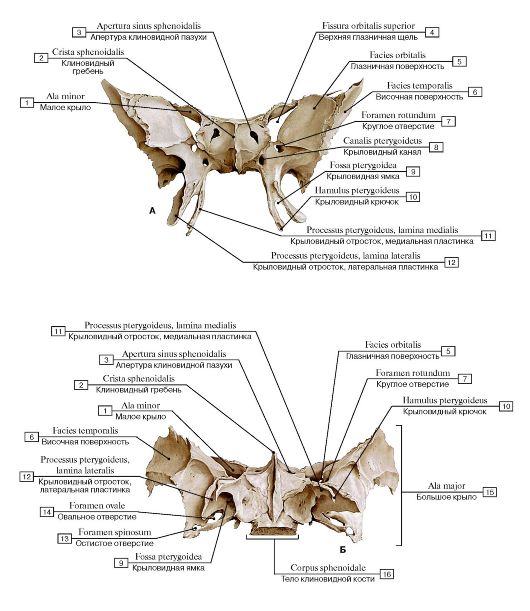 Рис. 43-1. Клиновидная кость (А - вид спереди, Б - вид снизу):
1 - Lesser wing; 2 - Sphenoidal crest; 3 - Opening of sphenoidal sinus; 4 - Superior orbital fissure; 5 - Orbital surface; 6 - Temporal surface; 7 - Foramen rotundum; 8 - Pterygoid canal; 9 - Pterygoid fossa; 10 - Pterygoid hamulus; 11 - Pterygoid process, medial plate; 12 - Pterygoid process, lateral plate; 13 - Foramen spinosum; 14 - Foramen ovale; 15 - Greater wing; 16 - Body of sphenoid
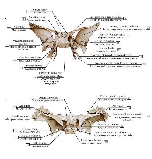 Рис. 43-2. Клиновидная кость (В - вид сзади, Г - вид сверху):
1 - Spongy bone; Trabecular bone; 2 - Pterygoid fossa; 3 - Pterygoid canal; 4 - Anterior clinoid process; 5 - Lesser wing; 6 - Optic canal; 7 - Dorsum sellae; 8 - Posterior clinoid process; 9 - Greater wing, cerebral surface; 10 - Superior orbital fissure; 11 - Foramen rotundum; 12 - Scaphoid fossa; 13 - Pterygoid process, lateral plate; 14 - Pterygoid process, medial plate; 15 - Sella turcica; 16 - Foramen spinosum; 17 - Foramen ovale; 18 - Jugum sphenoidale; Sphenoidal yoke; 19 - Greater wing; 20 - Hypophysial fossa
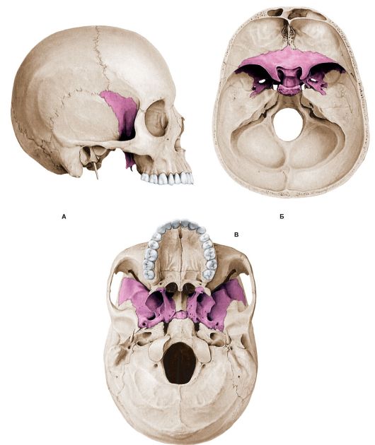 Рис. 44. Клиновидная кость в составе черепа (А - вид сбоку, справа, Б - вид сверху, В - вид снизу)
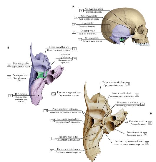 Рис.
45. Височная кость, правая (А - височная кость в составе черепа и ее
части - выделена цветом, Б - вид снизу, части височной кости выделены
разными цветами, В - вид снизу):
1 - Occipital bone; 2 - Temporal bone; 3 - Parietal bone; 4 - Sphenoid; Sphenoidal bone; 5 - Zygomatic bone; 6 - Petrous part; 7 - Squamous part; 8 - Tympanic part; 9 - Mandibular fossa; 10 - Styloid process; 11 - Mastoid foramen; 12 - Mastoid notch; 13 - Mastoid process; 14 - External acoustic opening; 15 - Zygomatic process; 16 - Articular tubercle; 17 - Carotid canal; 18 - Jugular fossa; 19 - Stylomastoid
foramen
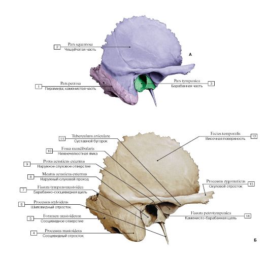 Рис. 46. Височная кость, правая (А - вид сбоку: части височной кости выделены разными цветами, Б - вид сбоку):
1 - Petrous part; 2 - Squamous part; 3 - Tympanic part; 4 - Mastoid process; 5 - Mastoid foramen; 6 - Styloid process; 7 - Tympanomastoid fissure; 8 - External acoustic meatus; 9 - External acoustic opening; 10 - Mandibular fossa; 11 - Articular tubercle; 12 - Temporal surface; 13 - Zygomatic process; 14 - Petrotympanic fissure
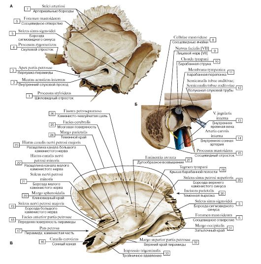 Рис. 47. Височная кость, правая (А - вид изнутри, Б - сообщения височной кости, В - вид изнутри и сверху):
1 - Styloid process; 2 - Internal acoustic meatus; 3 - Apex of petrous part; 4 - Zygomatic process; 5 - Groove for sigmoid sinus; 6 - Mastoid foramen; 7 - Arterial grooves; 8 - Mastoid cells; 9 - Facial nerve [VII]; 10 - Chorda tympani; 11 - Tympanic membrane; 12 - Canal for pharyngotympanic tube; Canal for auditory tube; 13 - Internaljugular vein; 14 - Internal carotid artery; 15 - Mastoid process; 16 - Carotid canal; 17 - Petrous part; 18 - Anterior surface of petrous part; 19 - Groove for greater petrosal nerve; 20 - Sphenoidal margin; 21 - Groove for lesser petrosal nerve; 22 - Hiatus for lesser petrosal nerve; 23 - Hiatus for greater petrosal nerve; 24 - Parietal margin; 25 - Cerebral surface; 26 - Petrosquamous fissure; 27 - Arcuate eminence; 28 - Tegmen tympani; 29 - Groove for superior petrosal sinus; 30 - Parietal notch; 31 - Occipital margin; 32 - Superior border of petrous part; 33 - Trigeminal impression
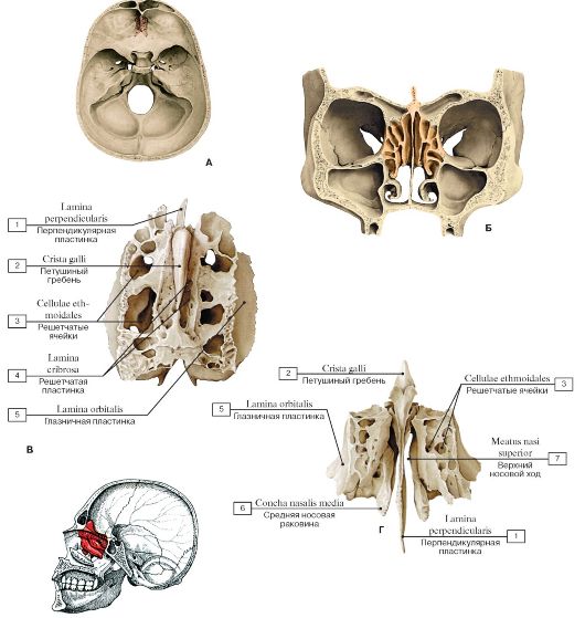 Д
Д
Рис. 48-1. Решетчатая кость (А - решетчатая кость в составе черепа, Б - положение решетчатой кости в лицевом черепе - фронтальное сечение через глазницы и носовую полость, В - вид сверху, Г - вид спереди, Д - топография
решетчатой кости):
1 - Perpendicular plate; 2 - Crista galli; 3 - Ethmoidal cells; 4 - Cribriform plate; 5 - Orbital plate; 6 - Middle nasal concha; 7 - Superior
nasal meatus
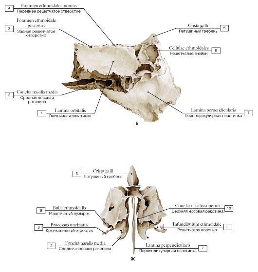 Рис. 48-2. Решетчатая кость (Е - вид сбоку, слева, Ж - вид сзади):
1 - Orbital plate; 2 - Middle nasal concha; 3 - Posterior ethmoidal foramen; 4 - Anterior ethmoidal foramen; 5 - Crista galli; 6 - Ethmoidal cells; 7 - Perpendicular plate; 8 - Uncinate process; 9 - Ethmoidal bulla; 10 - Superior nasal concha; 11 - Ethmoidal infundibulum
 Рис. 49. Нижняя носовая раковина, правая ( А - вид с медиальной стороны, Б - вид с латеральной стороны):
1 - Lacrimal process; 2 - Ethmoidal process; 3 - Maxillary process
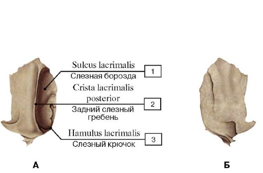 Рис. 50. Слезная кость, правая (А - вид снаружи, со стороны глазницы; Б - вид изнутри):
1 - Lacrimal groove; 2 - Posterior lacrimal crest; 3 - Lacrimal hamulus
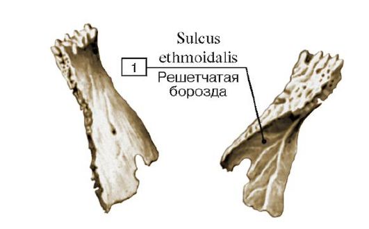 Рис. 51. Носовая кость, правая (А - вид снаружи, Б - вид изнутри):
1 - Ethmoidal groove
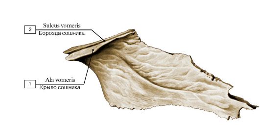
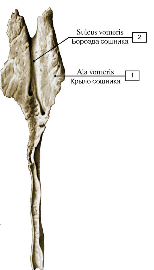 Рис. 52. Сошник ( А - вид справа, Б - вид сверху):
1 - Ala of vomer; 2 - Vomerine groove
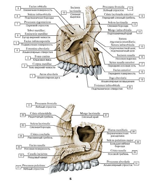 Рис. 53. Верхняя челюсть, правая (А - вид сбоку, с латеральной стороны, Б - вид с медиальной стороны):
1 - Alveolar arch; 2 - Body of maxilla; 3 - Canine fossa; 4 - Alveolar foramina; 5 - Infratemporal surface; 6 - Maxillary tuberosity; 7 - Zygomatic process; 8 - Infra-orbital groove; 9 - Orbital surface; 10 - Lacrimal notch; 11 - Frontal process; 12 - Anterior lacrimal crest; 13 - Lacrimal groove; 14 - Infra-orbital margin; 15 - Zygomaticomaxillary suture; 16 - Nasal notch; 17 - Anterior nasal spine; 18 - Anterior surface; 19 - Alveolar yokes; 20 - Infra-orbital foramen; 21 - Palatine process; 22 - Incisive canal; 23 - Nasal surface; 24 - Conchal crest; 25 - Lacrimal groove; 26 - Ethmoidal crest; 27 - Lacrimal margin; 28 - Maxillary hiatus; 29 - Greater palatine groove; 30 - Nasal
crest; 31 - Alveolar process
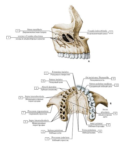 Рис. 54. Верхняя челюсть, правая (А - вид с латеральной стороны, Б - верхние челюсти, вид снизу):
1 - зонды в Alveolar canals; 2 - Maxillary sinus; 3 - Infra-orbital canal; 4 - Palatine process; 5 - Palatine spines; 6 - Interradicular septa; 7 - Zygomatic process; 8 - Interalveolar septa; 9 - Dental alveoli; 10 - Incisive suture; 11 - Incisive foramina; 12 - Incisive bone; Premaxilla; 13 - Median palatine suture; 14 - Alveolar arch; 15 - Palatine grooves; 16 - Palatine torus
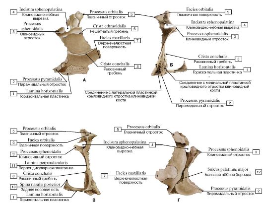
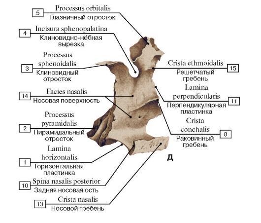 Рис. 55. Нёбная кость, левая (А - вид изнутри,
медиальная сторона, Б - вид сзади, справа, В - вид спереди, Г - вид снаружи, латеральная сторона, Д - вид сзади и изнутри):
1 - Horizontal plate; 2 - Pyramidal process; 3 - Sphenoidal process; 4 - Sphenopalatine notch; 5 - Orbital process; 6 - Ethmoidal crest; 7 - Maxillary surface; 8 - Conchal crest; 9 - Orbital surface; 10 - Posterior nasal spine; 11 - Perpendicular plate; 12 - Greater palatine groove; 13 - Nasal crest; 14 - Nasal surface; 15 - Ethmoidal crest
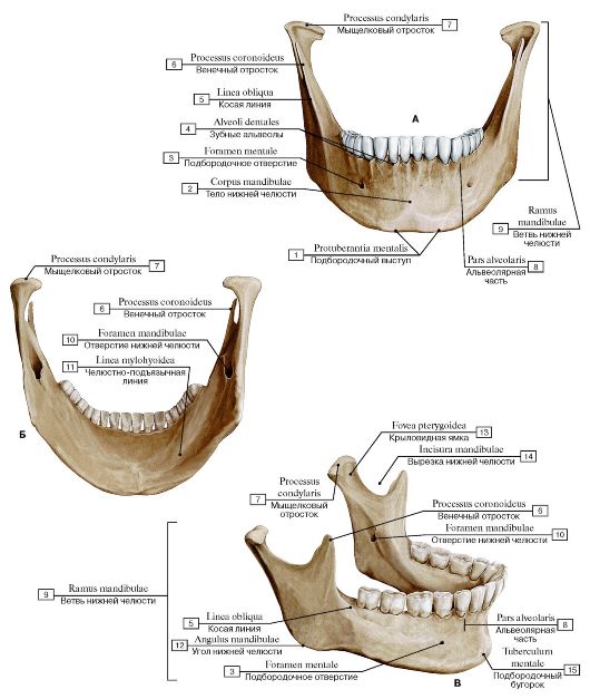 Рис. 56. Нижняя челюсть (А - вид спереди, Б - вид сзади, В - вид сбоку, справа):
1 - Mental protuberance; 2 - Body of mandible; 3 - Mental foramen; 4 - Dental alveoli; 5 - Oblique line; 6 - Coronoid process; 7 - Condylar process; 8 - Alveolar part; 9 - Ramus of mandible; 10 - Mandibular foramen; 11 - Mylohyoid line; 12 - Angle of mandible; 13 - Pterygoid fovea; 14 - Mandibular notch; 15 - Mental tubercle
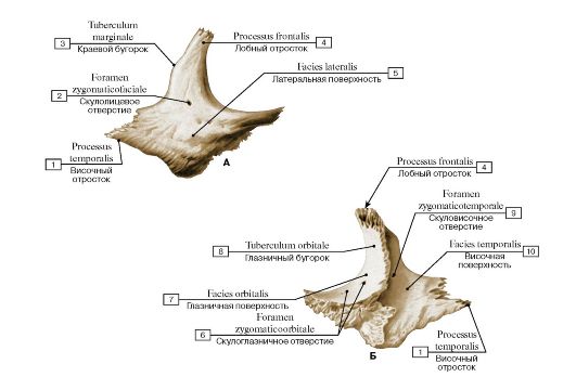 Рис. 57. Скуловая кость, правая (А - вид снаружи, Б - вид изнутри):
1 - Temporal process; 2 - Zygomaticofacial foramen; 3 - Marginal tubercle; 4 - Frontal process; 5 - Lateral surface; 6 - Zygomatico-orbital foramen; 7 - Orbital surface; 8 - Orbital tubercle; 9 - Zygomaticotemporal foramen; 10 - Temporal surface
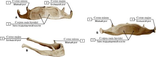 Рис. 58. Подъязычная кость (А - вид спереди, Б - вид сзади, В - вид сбоку):
1 - Lesser horn; 2 - Greater horn; 3 - Body of hyoid bone
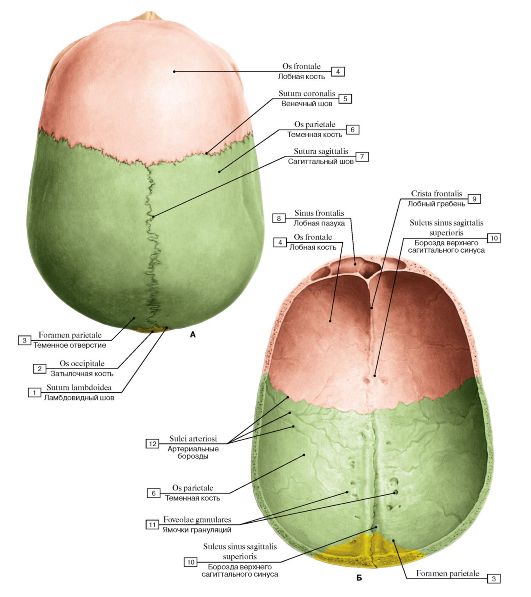 Теменное отверстие
Рис. 59. Свод (крыша) черепа (А - вид сверху, Б - вид изнутри, со стороны полости черепа):
1 - Lambdoid suture; 2 - Occipital bone; 3 - Parietal foramen; 4 - Frontal bone; 5 - Coronal suture; 6 - Parietal bone; 7 - Sagittal suture; 8 - Frontal sinus; 9 - Frontal crest; 10 - Groove for superior sagittal sinus; 11 - Granular foveolae; 12 - Arterial grooves
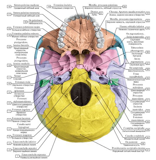 Рис. 60. Наружное основание черепа:
1 - Highest nuchal line; 2 - Superior nuchal line; 3 - Inferior nuchal line; 4 - Foramen magnum; 5 - Hypoglossal canal; 6 - Foramen lacerum; 7 - Jugular foramen; 8 - Stylomastoid foramen; 9 - Foramen spinosum; 10 - Foramen ovale; 11 - Vomer; 12 - Pterygoid process, medial plate; 13 - Pterygoid process, lateral plate; 14 - Lesser palatine foramina; 15 - Greater palatine foramen; 16 - Palatine bone; 17 - Transverse palatine suture; 18 - Median palatine suture; 19 - Incisive foramina; 20 - Maxilla, palatine process; 21 - Teeth; 22 - Choana; Posterior nasal aperture; 23 - Maxilla, zygomatic process; 24 - Inferior orbital fissure; 25 - Zygomatic bone, temporal surface; 26 - Pharyngeal tubercle; 27 - Zygomatic arch; 28 - Temporal bone; 29 - Mandibular fossa; 30 - Styloid process; 31 - Mastoid process; 32 - Mastoid notch; 33 - Mastoid foramen; 34 - Occipital condyle; 35 - Condylar canal; 36 - Parietal bone; 37 - External occipital protuberance
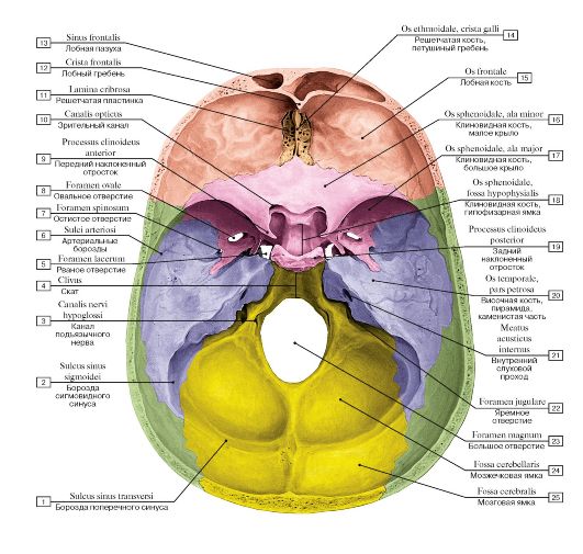 Рис. 61. Внутреннее основание черепа:
1 - Groove for transverse sinus; 2 - Groove for sigmoid sinus; 3 - Hypoglossal canal; 4 - Clivus; 5 - Foramen lacerum; 6 - Arterial grooves; 7 - Foramen spinosum; 8 - Foramen ovale; 9 - Anterior clinoid process; 10 - Optic canal; 11 - Cribriform plate; 12 - Frontal crest; 13 - Frontal sinus; 14 - Ethmoid; Ethmoidal bone, crista galli; 15 - Frontal bone; 16 - Sphenoid; Sphenoidal bone, lesser wing; 17 - Sphenoid; Sphenoidal bone, greater wing; 18 - Sphenoid; Sphenoidal bone, hypophysial fossa; 19 - Posterior clinoid process; 20 - Temporal bone, petrous part; 21 - Internal acoustic meatus; 22 - Jugular foramen; 23 - Foramen magnum; 24 - Cerebellar fossa; 25 - Cerebral
fossa
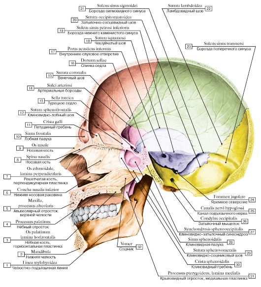 Рис. 62. Череп, вид изнутри, сбоку:
1 - Mylohyoid line; 2 - Mandible; 3 - Palatine bone, horizontal plate; 4 - Palatine process; 5 - Maxilla, alveolar process; 6 - Inferior nasal concha; 7 - Ethmoid; Ethmoidal bone, perpendicular plate; 8 - Nasal spine; 9 - Nasal bone; 10 - Frontal sinus; 11 - Crista galli; 12 - Sphenofrontal suture; 13 - Sella turcica; 14 - Arterial grooves; 15 - Coronal suture; 16 - Dorsum sellae; 17 - Internal acoustic opening; 18 - Squamous suture; 19 - Groove for inferior petrosal sinus; 20 - Occipitomastoid suture; 21 - Groove for sigmoid sinus; 22 - Lambdoid suture; 23 - Groove for transverse sinus; 24 - Jugular foramen; 25 - Hypoglossal canal; 26 - Occipital condyle; 27 - Spheno-occipital synchondrosis; 28 - Sphenoidal sinus; 29 - Sphenovomerine suture; 30 - Sphenoidal crest; 31 - Pterygoid process, medial plate; 32 - Vomer
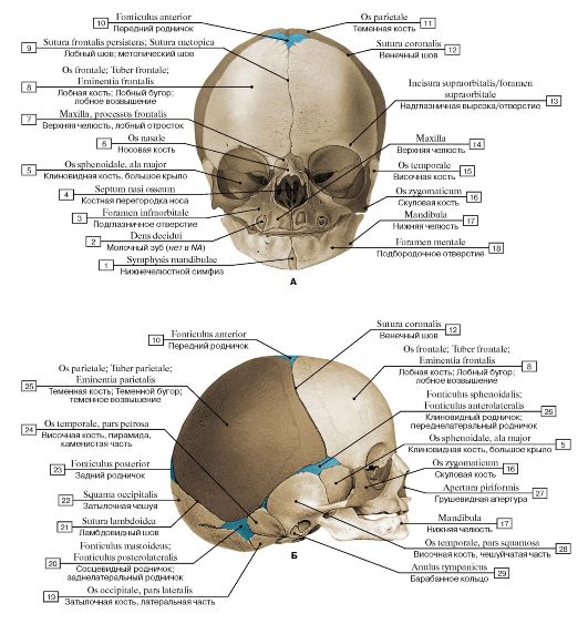 Рис. 63. Череп новорожденного, роднички (А - вид спереди, Б - вид сбоку, справа):
1 - Mandibtilar symphysis; 2 - Milk tooth; 3 - Infra-orbital foramen; 4 - Bony nasal septum; 5 - Sphenoid; Sphenoidal bone, greater wing; 6 - Nasal bone; 7 - Maxilla, frontal process; 8 - Frontal bone; Frontal tuber; Frontal eminence; 9 - Frontal suture; Metopic suture; 10 - Anterior fontanelle; 11 - Parietal bone; 12 - Coronal suture; 13 - Supra-orbital notch/foramen; 14 - Maxilla; 15 - Temporal bone; 16 - Zygomatic bone; 17 - Mandible; 18 - Mental foramen; 19 - Occipital bone, lateral part; 20 - Mastoid fontanelle; 21 - Lambdoid suture; 22 - Squamous part of occipital bone; 23 - Posterior fontanelle; 24 - Temporal bone, petrous part; 25 - Parietal bone; Parietal tuber; Parietal eminence; 26 - Sphenoidal fontanelle; 27 - Piriform aperture; 28 - Temporal bone, squamous part; 29 - Tympanic ring
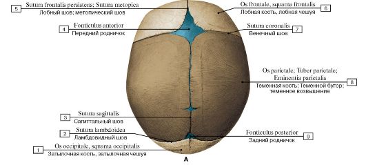
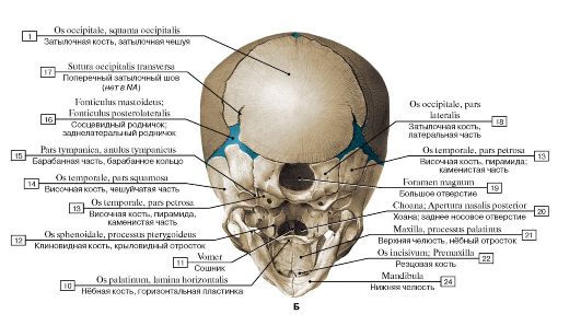 Рис. 64. Череп новорожденного, роднички (А - вид сверху, Б - вид снизу):
1 - Occipital bone, squamous part of occipital bone; 2 - Lambdoid suture; 3 - Sagittal suture; 4 - Anterior fontanelle; 5 - Frontal suture; Metopic suture; 6 - Frontal bone; squamous part; 7 - Coronal suture; 8 - Parietal bone; Parietal tuber; Parietal eminence; 9 - Posterior fontanelle; 10 - Palatine bone, horizontal plate; 11 - Vomer; 12 - Sphenoid; Sphenoidal bone, pterygoid process; 13 - Temporal bone, petrous part; 14 - Temporal bone, squamous part; 15 - Tympanic part, tympanic ring; 16 - Mastoid fontanelle; 17 - Transverse occipital suture; 18 - Occipital bone, lateral part; 19 - Foramen magnum; 20 - Choana; Posterior nasal aperture; 21 - Maxilla, palatine process;
22 - Incisive bone; Premaxilla; 23 - Mandible
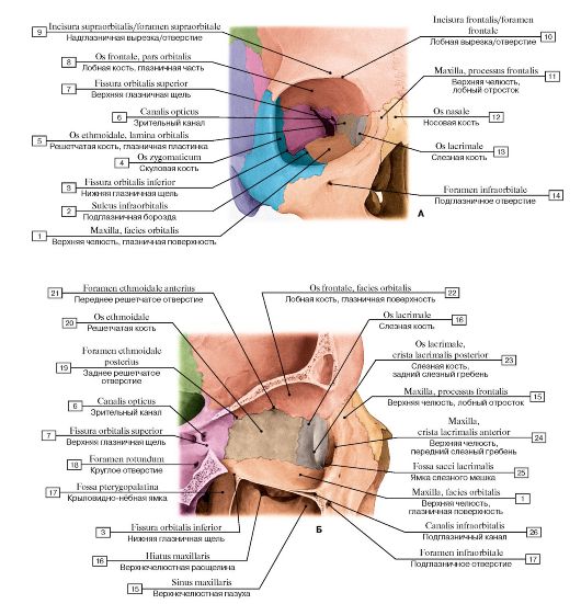 Рис. 65. Глазница, правая (А - вид спереди, Б - вид сбоку снаружи, распил проходит по глазнице, видна медиальная стенка):
1 - Maxilla, orbital surface; 2 - Infra-orbital groove; 3 - Inferior orbital fissure; 4 - Zygomatic bone; 5 - Ethmoid; Ethmoidal bone, orbital plate; 6 - Optic canal; 7 - Superior orbital fissure; 8 - Frontal bone, orbital part; 9 - Supra-orbital notch/foramen; 10 - Frontal notch/foramen; 11 - Maxilla, frontal process; 12 - Nasal bone; 13 - Lacrimal bone; 14 - Infra-orbital foramen; 15 - Maxillary sinus; 16 - Maxillary hiatus; 17 - Pterygopalatine fossa; 18 - Foramen rotundum; 19 - Posterior ethmoidal foramen; 20 - Ethmoid; Ethmoidal bone; 21 - Anterior ethmoidal foramen; 22 - Frontal bone, orbital surface; 23 - Lacrimal bone, posterior lacrimal crest; 24 - Maxilla, anterior lacrimal crest; 25 - Fossa for lacrimal sac; 27 - Infra-orbital canal
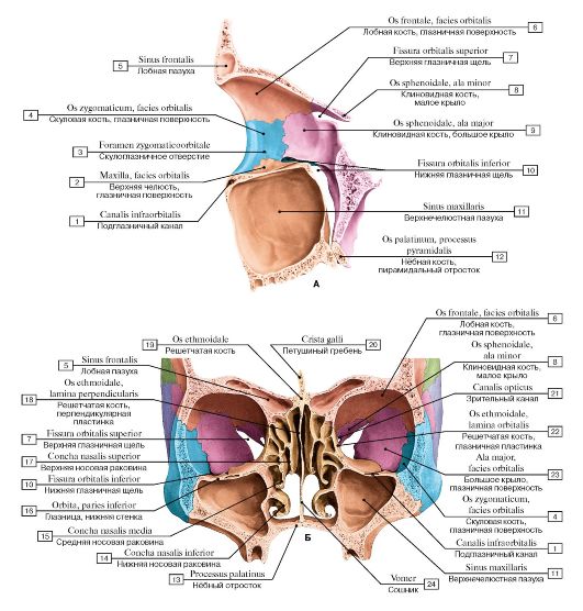 Рис.
66. Глазница, левая (А - вид сбоку изнутри, распил проходит по
глазнице, видна латеральная стенка, Б - глазницы и носовая полость с
окружающими воздухоносными полостями (пазухами) черепа):
1 - Infra-orbital canal; 2 - Maxilla, orbital surface; 3 - Zygomatico-orbital foramen; 4 - Zygomatic bone, orbital surface; 5 - Frontal sinus; 6 - Frontal bone, orbital surface; 7 - Superior orbital fissure; 8 - Sphenoid; Sphenoidal bone, lesser wing; 9 - Sphenoid; Sphenoidal bone, greater wing; 10 - Inferior orbital fissure; 11 - Maxillary sinus; 12 - Palatine bone, pyramidal process; 13 - Palatine process; 14 - Inferior nasal concha; 15 - Middle nasal concha; 16 - Orbit, floor; 17 - Superior nasal concha; 18 - Ethmoid; Ethmoidal bone, perpendicular plate; 19 - Ethmoid; Ethmoidal bone; 20 - Crista galli; 21 - Optic canal; 22 - Ethmoid; Ethmoidal bone, orbital plate; 23 - Greater wing, orbital
surface; 24 - Vomer
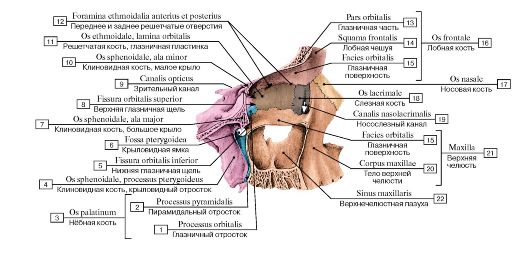 Рис. 67. Медиальная стенка глазницы, правой, вид сбоку:
1 - Orbital process; 2 - Pyramidal process; 3 = 1 + 2 - Palatine bone; 4 - Sphenoid; Sphenoidal bone, pterygoid process; 5 - Inferior orbital fissure; 6 - Pterygoid fossa; 7 - Sphenoid; Sphenoidal bone, greater wing; 8 - Superior orbital fissure; 9 - Optic canal; 10 - Sphenoid; Sphenoidal bone, lesser wing; 11 - Ethmoid; Ethmoidal bone, orbital plate; 12 - Anterior and posterior ethmoidal foramen; 13 - Orbital part; 14 - Squamous part; 15 - Orbital surface; 16 = 13 + 14 + 15 - Frontal bone; 17 - Nasal bone; 18 - Lacrimal bone; 19 - Nasolacrimal canal; 20 - Body of maxilla; 21 = 15 + 20 - Maxilla; 22 - Maxillary sinus
 АБ
АБ
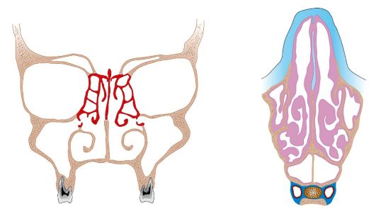
Рис. 68. Околоносовые пазухи (А - фронтальный разрез, Б - поперечный разрез)
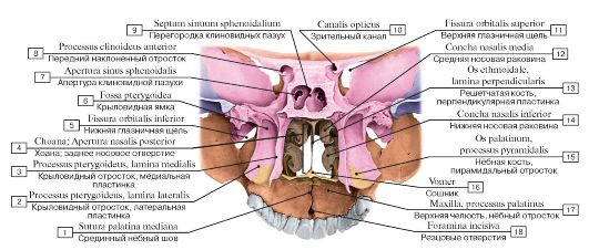 Рис. 69. Скелет полости носа и твердого нёба, вид сзади:
1 - Median palatine suture; 2 - Pterygoid process, lateral plate; 3 - Pterygoid process, medial plate; 4 - Choana; Posterior nasal aperture; 5 - Inferior orbital fissure; 6 - Pterygoid fossa; 7 - Opening of sphenoidal sinus; 8 - Anterior clinoid process; 9 - Septum of sphenoidal sinuses; 10 - Optic canal; 11 - Superior orbital fissure; 12 - Middle nasal concha; 13 - Ethmoid; Ethmoidal bone, perpendicular plate; 14 - Inferior nasal concha; 15 - Palatine bone, pyramidal process; 16 - Vomer; 17 - Maxilla, palatine process; 18 - Incisive foramina
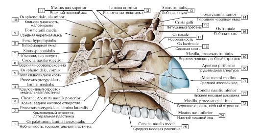 Рис. 70. Боковая (латеральная) стенка полости носа, левая:
1 - Palatine bone, horizontal plate; 2 - Pterygoid process, lateral plate; 3 - Choana; Posterior nasal aperture; 4 - Pterygoid process, medial plate; 5 - Sphenoid; Sphenoidal bone, body; 6 - Superior nasal concha; 7 - Sphenoidal sinus; 8 - Hypophysial fossa; 9 - Middle cranial fossa; 10 - Sphenoid; Sphenoidal bone, lesser wing; 11 - Superior nasal meatus; 12 - Cribriform plate; 13 - Frontal sinus; 14 - Anterior cranial fossa; 15 - Crista galli; 16 - Frontal bone; 17 - Nasal bone; 18 - Lacrimal bone; 19 - Maxilla, frontal process; 20 - Piriform aperture; 21 - Middle nasal meatus; 22 - Inferior nasal concha; 23 - Maxilla, palatine process; 24 - Inferior nasal meatus; 25 - Middle
nasal concha
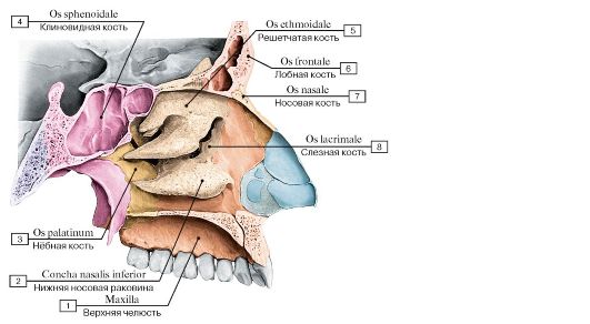 Рис. 71. Боковая стенка полости носа, левая:
1 - Maxilla; 2 - Inferior nasal concha; 3 - Palatine bone; 4 - Sphenoid; Sphenoidal bone; 5 - Ethmoid; Ethmoidal bone; 6 - Frontal bone; 7 - Nasal bone; 8 - Lacrimal bone
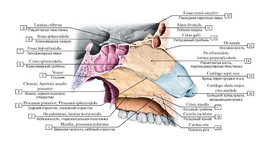 Рис. 72. Костная перегородка носа, вид справа:
I - Maxilla, palatine process; 2 - Palatine bone, horizontal plate; 3 - Posterior process; Sphenoid process; 4 - Choana; Posterior nasal aperture; 5 - Vomer; 6 - Sphenoidal crest; 7 - Hypophysial fossa; 8 - Sphenoidal sinus; 9 - Cribriform plate; 10 - Anterior cranial fossa;
II - Frontal sinus; 12 - Crista galli; 13 - Nasal bone; 14 - Ethmoid; Ethmoidal bone, perpendicular plate; 15 - Septal nasal cartilage;
16 - Major alar cartilage, medial crus; 17 - Nasal crest; 18 - Incisive canal; 19 - Oral cavity
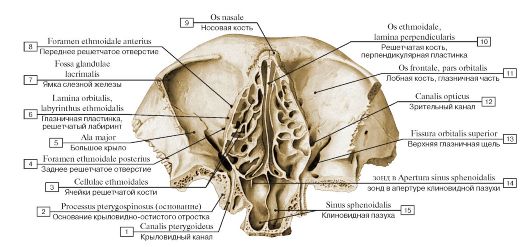 Рис. 73. Скелет полости носа и глазниц, вид снизу (горизонтальный распил через срединные отделы входа в глазницы):
1 - Pterygoid canal; 2 - Pterygospinous process; 3 - Ethmoidal cells; 4 - Posterior ethmoidal foramen; 5 - Greater wing; 6 - Orbital plate, ethmoidal labyrinth; 7 - Fossa for lacrimal gland; Lacrimal fossa; 8 - Anterior ethmoidal foramen; 9 - Nasal bone; 10 - Ethmoid; Ethmoidal bone, perpendicular plate; 11 - Frontal bone, orbital part; 12 - Optic canal; 13 - Superior orbital fissure; 14 - зонд в Opening of
sphenoidal sinus; 15 - Sphenoidal sinus
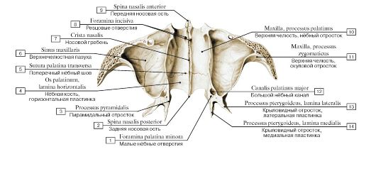 Рис. 74. Нижняя стенка полости носа (костное нёбо), вид сверху (горизонтальный распил через скуловые отростки верхних челюстей):
1 - Lesser palatine foramina; 2 - Posterior nasal spine; 3 - Pyramidal process; 4 - Palatine bone, horizontal plate; 5 - Transverse palatine suture; 6 - Maxillary sinus; 7 - Nasal crest; 8 - Incisive foramina; 9 - Anterior nasal spine; 10 - Maxilla , palatine process; 11 - Maxilla, zygomatic process; 12 - Greater palatine canal; 13 - Pterygoid process, lateral plate; 14 - Pterygoid process, medial plate
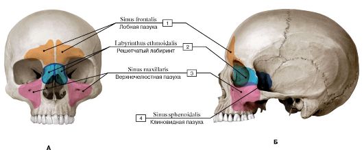
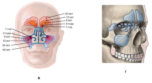 Рис.
75. Пазухи воздухоносных костей черепа (околоносовые пазухи) (выделены
цветом) (А - вид спереди, Б - вид сбоку, слева, В - возрастные изменения
лобной и верхнечелюстной пазух, Г - проекции воздухоносных пазух
черепа):
1 - Frontal sinus; 2 - Ethmoidal labyrinth; 3 - Maxillary sinus; 4 - Sphenoidal sinus
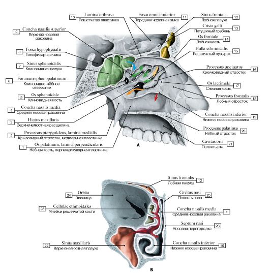 Рис. 76. Полость носа (А - латеральная (левая) стенка, вид справа, Б - полость носа и правая глазница):
1 - Palatine bone, perpendicular plate; 2 - Pterygoid process, medial plate; 3 - Maxillary hiatus; 4 - Middle nasal concha; 5 - Sphenoid; Sphenoidal bone; 6 - Sphenopalatine foramen; 7 - Sphenoidal sinus; 8 - Hypophysial fossa; 9 - Superior nasal concha; 10 - Cribriform plate; 11 - Anterior cranial fossa; 12 - Frontal sinus; 13 - Crista galli; 14 - Frontal bone; 15 - Ethmoidal bulla; 16 - Uncinate process; 17 - Lacrimal bone; 18 - Frontal process; 19 - Inferior nasal concha; 20 - Palatine process; 21 - Oral cavity; 22 - Maxillary sinus; 23 - Ethmoidal cells; 24 - Orbit; 25 - Nasal cavity; 26 - Nasal septum
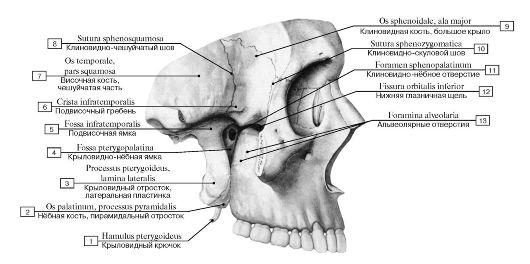 Рис. 77. Височная ямка, подвисочная ямка и крыловидно-нёбная ямка, вид справа, скуловая дуга удалена:
1 - Pterygoid hamulus; 2 - Palatine bone, pyramidal process; 3 - Pterygoid process, lateral plate; 4 - Pterygopalatine fossa; 5 - Infratemporal fossa; 6 - Infratemporal crest; 7 - Temporal bone, squamous part; 8 - Sphenosquamous suture; 9 - Sphenoid; Sphenoidal bone, greater wing; 10 - Sphenozygomatic suture; 11 - Sphenopalatine foramen; 12 - Inferior orbital fissure;
13 - Alveolar foramina
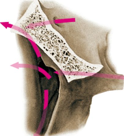 Рис.
78. Крыловидно-нёбная ямка, вид снизу. Стрелки, изображенные на схеме,
показывают доступ к крыловидно- нёбной ямке через основание черепа. Сама
ямка (на рисунке не показана) расположена сбоку от латеральной
пластинки
крыловидного отростка клиновидной кости
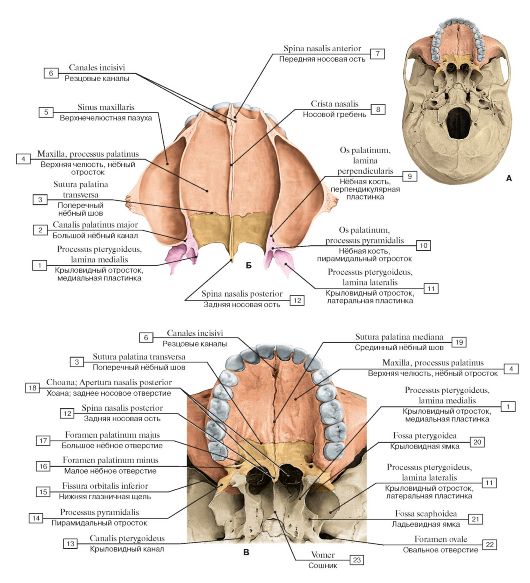 Рис. 79. Твердое нёбо (А - положение твердого нёба на черепе, вид снизу, Б - вид сверху, В - вид снизу):
1 - Pterygoid process, medial plate; 2 - Greater palatine canal; 3 - Transverse palatine suture; 4 - Maxilla, palatine process; 5 - Maxillary sinus; 6 - Incisive canals; 7 - Anterior nasal spine; 8 - Nasal crest; 9 - Palatine bone, perpendicular plate; 10 - Palatine bone, pyramidal process; 11 - Pterygoid process, lateral plate; 12 - Posterior nasal spine; 13 - Pterygoid canal; 14 - Pyramidal process; 15 - Inferior orbital fissure; 16 - Lesser palatine foramen; 17 - Greater palatine foramen; 18 - Choana; Posterior nasal aperture; 19 - Median palatine suture; 20 - Pterygoid fossa; 21 - Scaphoid fossa; 22 - Foramen ovale; 23 - Vomer
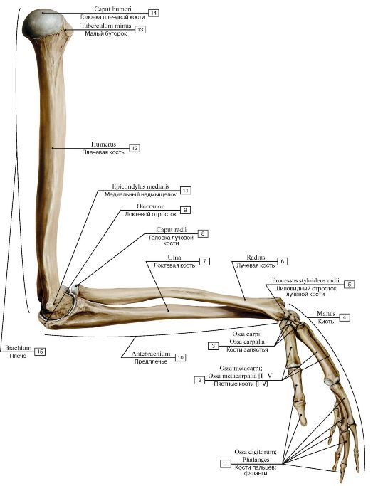 Рис. 80. Кости верхней конечности, левой, вид сбоку:
1 - Phalanges; 2 - Metacarpals [I-V]; 3 - Carpal bones; 4 - Hand; 5 - Radial styloid process; 6 - Radius; 7 - Ulna; 8 - Head of radius; 9 - Olecranon; 10 - Forearm; 11 - Medial epicondyle; 12 - Humerus; 13 - Lesser tubercle; 14 - Head of humerus; 15 - Arm
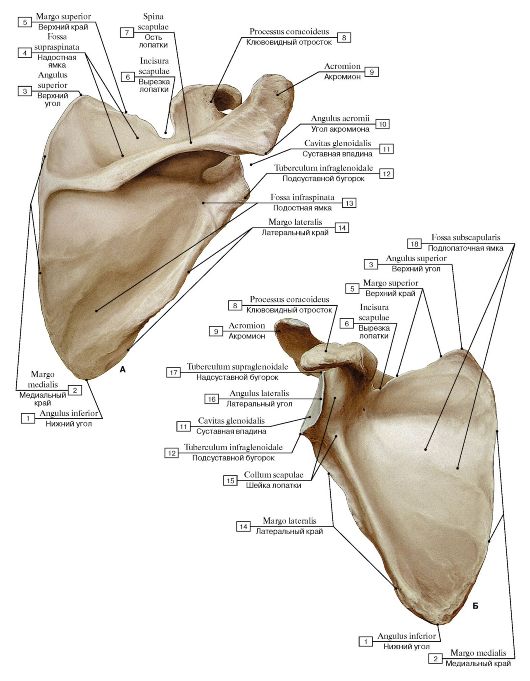 Рис. 81. Лопатка, правая (А - вид спереди, Б - вид сзади):
1 - Inferior angle; 2 - Medial border; 3 - Superior angle; 4 - Supraspinous fossa; 5 - Superior border; 6 - Suprascapular notch; 7 - Spine of scapula; 8 - Coracoid process; 9 - Acromion; 10 - Acromial angle; 11 - Glenoid cavity; 12 - Infraglenoid tubercle; 13 - Infraspinous fossa; 14 - Lateral border; 15 - Neck of scapula; 16 - Lateral angle; 17 - Supraglenoid tubercle; 18 - Subscapular fossa
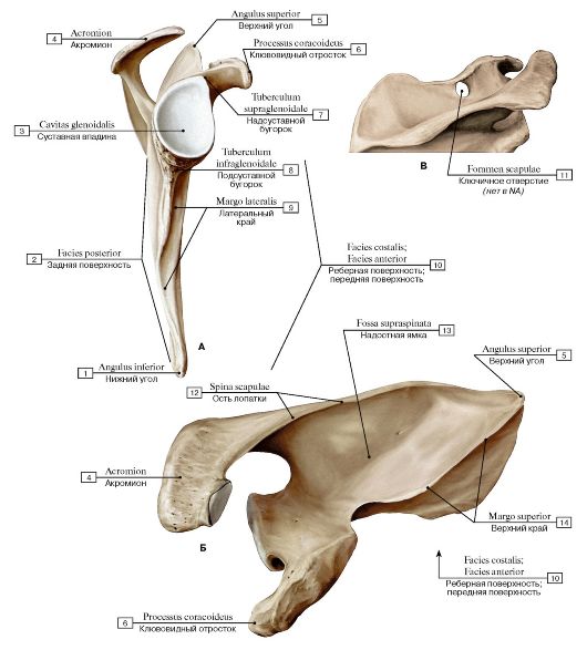 Рис.
82. Лопатка, правая (А - вид с латеральной стороны, Б - вид сверху, В -
лопаточное отверстие, анатомический вариант, вид сверху):
1 - Inferior angle; 2 - Posterior surface; 3 - Glenoid cavity; 4 - Acromion; 5 - Superior angle; 6 - Coracoid process; 7 - Supraglenoid tubercle; 8 - Infraglenoid tubercle; 9 - Lateral border; 10 - Costal surface; 11 - Scapular foramen; 12 - Spine of scapula; 13 - Supraspinous
fossa; 14 - Superior border
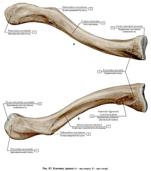 1
- Acromial end; 2 - Conoid tubercle; 3 - Shaft of clavicle; Body of
clavicle; 4 - Sternal facet; 5 - Sternal end; 6 - Impression for
costoclavicular ligament; 7 - Subclavian groove; Groove for subclavius; 8
- Acromial facet
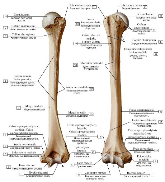 Рис. 84. Плечевая кость, правая (А - вид спереди, Б - вид сзади):
1 - Trochlea; 2 - Olecranon fossa; 3 - Medial epicondyle; 4 - Groove for ulnar nerve; 5 - Medial supraepicondylar ridge; Medial supracondylar ridge; 6 - Medial border; 7 - Shaft of humerus; Body of humerus, posterior surface; 8 - Surgical neck; 9 - Anatomical neck; 10 - Head of humerus; 11 - Greater tubercle; 12 - Radial groove; Groove for radial nerve; 13 - Lateral margin; 14 - Medial supraepicondylar ridge; Medial supracondylar ridge; 15 - Lateral epicondyle; 16 - Capitulum; 17 - Radial fossa; 18 - Deltoid tuberosity; 19 - Crest of greater tubercle; Lateral lip; 20 - Intertubercular sulcus; Bicipital groove; 21 - Lesser tubercle; 22 - Crest of lesser tubercle; Medial lip; 23 - Anteromedial surface; 24 - Anterolateral surface; 25 - Coronoid fossa
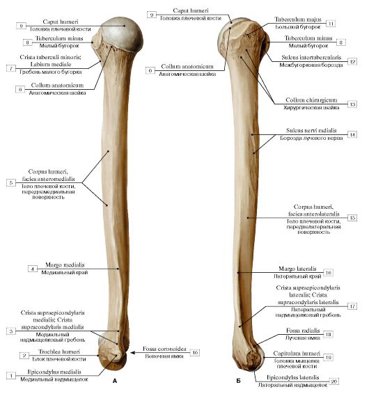 Рис. 85. Плечевая кость, правая (А - медиальная сторона, Б - латеральная сторона):
1 - Medial epicondyle; 2 - Trochlea; 3 - Medial supraepicondylar ridge; Medial supracondylar ridge; 4 - Medial border; 5 - Shaft of humerus; Body of humerus, anteromedial surface; 6 - Anatomical neck; 7 - Crest of lesser tubercle; Medial lip; 8 - Lesser tubercle; 9 - Head of humerus; 10 - Coronoid fossa; 11 - Greater tubercle; 12 - Intertubercular sulcus; Bicipital groove; 13 - Surgical neck; 14 - Radial groove; Groove for radial nerve; 15 - Shaft of humerus; Body of humerus, anterolateral surface; 16 - Lateral border; 17 - Lateral supraepicondylar ridge; Lateral supracondylar ridge; 18 - Radial fossa; 19 - Capitulum; 20 - Lateral epicondyle
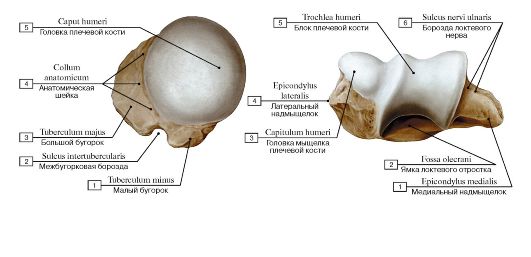 Рис. 86. Головка плечевой кости, правой:
1 - Lesser tubercle; 2 - Intertubercular sulcus; Bicipital groove; 3 - Greater tubercle; 4 - Anatomical neck; 5 - Head of humerus
Рис. 87. Мыщелок плечевой кости, правой:
1 - Medial epicondyle; 2 - Olecranon fossa; 3 - Capitulum; 4 - Lateral epicondyle; 5 - Trochlea; 6 - Groove for ulnar nerve
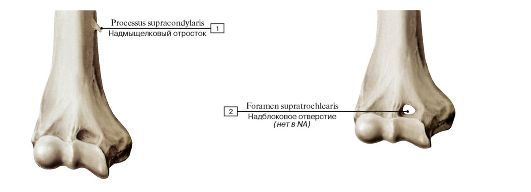 Рис. 88. Варианты развития дистального эпифиза плеча, правого, вид спереди:
1 - Supracondylar process; 2 - Supratrohlear foramen
 Рис. 89. Повреждения верхнего эпифиза плеча, правого, вид спереди:
1 - Intertubercular sulcus; Bicipital groove; 2 - Greater tubercle; 3 - Lesser tubercle; 4 - Surgical neck; 5 - Head of humerus; 6 - Anatomi-
cal neck
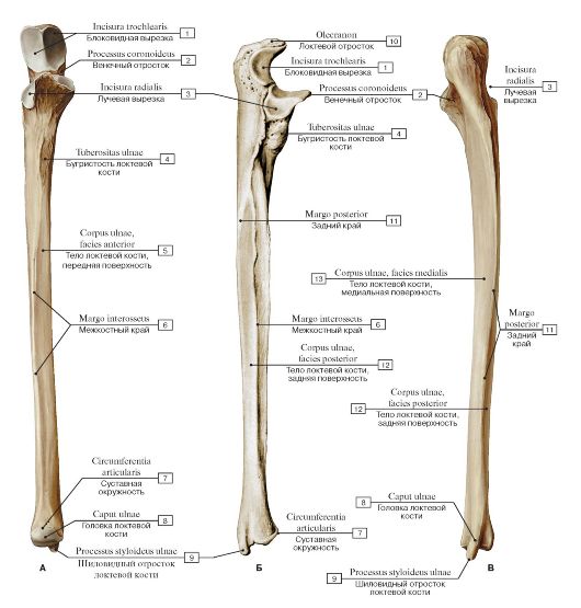 Рис. 90. Локтевая кость, правая (А - вид спереди, Б - вид с латеральной стороны, В - вид сзади):
1 - Trochlear notch; 2 - Coronoid process; 3 - Radial notch; 4 - Tuberosity of ulna; 5 - Shaft of ulna; Body of ulna, anterior surface; 6 - Interosseous border; 7 - Articular circumference; 8 - Head of ulna; 9 - Ulnar styloid process; 10 - Olecranon; 11 - Posterior border; 12 - Shaft of ulna; Body of ulna, posterior surface;13 - Shaft of ulna; Body of ulna, medial surface
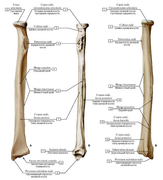 Рис. 91. Лучевая кость, правая (А - вид спереди, Б - вид с медиальной стороны, В - вид сзади):
1 - Head of radius; Articular circumference; 2 - Articular facet; 3 - Neck of radius; 4 - Radial tuberosity; 5 - Anterior border; 6 - Interosseous border; 7 - Shaft of radius; Body of radius, anterior surface; 8 - Carpal articular surface; 9 - Radial styloid process; 10 - Shaft of radius; Body of radius, posterior surface; 11 - Ulnar notch; 12 - Posterior border; 13 - Shaft of radius; Body of radius, lateral surface;
14 - Dorsal tubercle
 Локтевая кость
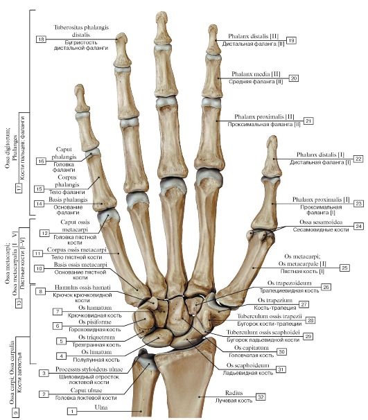
Рис. 92.
Кости кисти, правой, ладонная поверхность:
1 - Ulna; 2 - Head of ulna; 3 - Ulnar styloid process; 4 - Lunate; 5 - Triquetrum; 6 - Pisiform; 7 - Hamate; 8 - Hook of hamate; 9 - Carpal bones; 10 - Base of metacarpal; 11 - Shaft of metacarpal; Body of metacarpal; 12 - Head of metacarpal; 13 - Metacarpals [I-V]; 14 - Base of phalanx; 15 - Shaft of phalanx; Body of phalanx; 16 - Head of phalanx; 17 - Phalanges; 18 - Tuberosity of distal phalanx; 19 - Distal phalanx [II]; 20 - Middle phalanx [II]; 21 - Proximal phalanx [II]; 22 - Distal phalanx [I]; 23 - Proximal phalanx [I]; 24 - Sesamoid bones; 25 - Metacarpal [I]; 26 - Trapezoid; 27 - Trapezium; 28 - Trapezium, tubercle; 29 - Tubercle of scaphoid; 30 - Capitate; 31 - Scaphoid; 32 - Radius
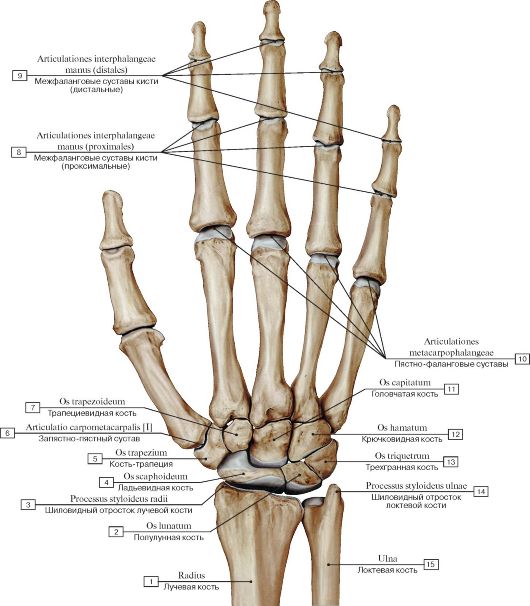 Рис. 93. Кости кисти, правой, тыльная сторона:
1 - Radius; 2 - Lunate; 3 - Radial styloid process; 4 - Scaphoid; 5 - Trapezium; 6 - Carpometacarpal joint [I]; 7 - Trapezoid; 8 - Interphalangeal joints of hand (proximal); 9 - Interphalangeal joints of hand (distal); 10 - Metacarpophalangeal joints; 11 - Capitate; 12 - Hamate; 13 - Triquetrum; 14 - Ulnar styloid process; 15 - Ulna
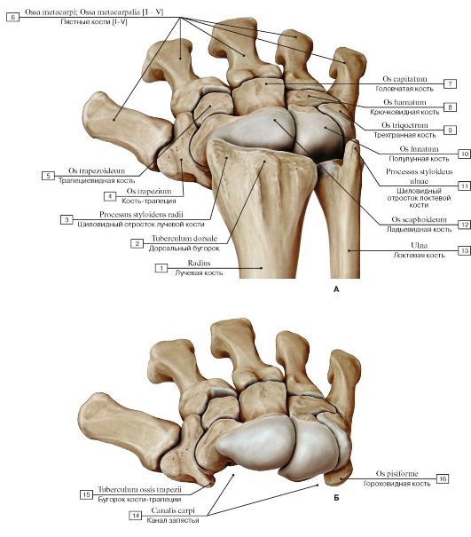 Рис.
94. Кости пясти и запястья, правых (А - дистальные эпифизы костей
предплечья и кости запястья, Б - вид кисти после удаления костей
предплечья):
1 - Radius; 2 - Dorsal tubercle; 3 - Radial styloid process; 4 - Trapezium; 5 - Trapezoid; 6 - Metacarpals [I-V]; 7 - Capitate; 8 - Hamate; 9 - Triquetrum; 10 - Lunate; 11 - Ulnar styloid process; 12 - Scaphoid; 13 - Ulna; 14 - Carpal tunnel; 15 - Trapezium, tubercle;
16 - Pisiform
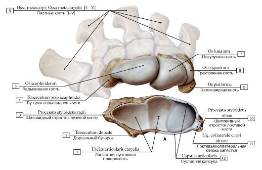
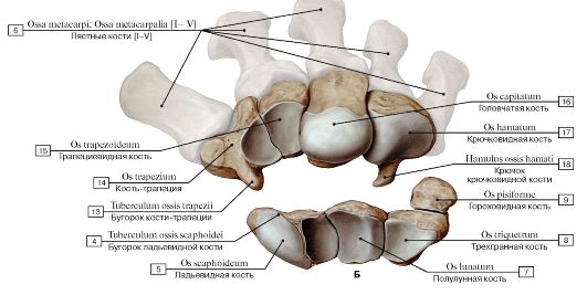 Рис. 95. Кости запястья, правого (А - проксимальный ряд, Б - дистальный ряд):
1 - Radius, carpal articular surface; 2 - Dorsal tubercle; 3 - Radial styloid process; 4 - Scaphoid, tubercle; 5 - Scaphoid; 6 - Metacarpals [I-V]; 7 - Lunate; 8 - Triquetrum; 9 - Pisiform; 10 - Ulnar styloid process; 11 - Ulnar collateral ligament of wrist joint; 12 - Joint capsule; Articular capsule; 13 - Trapezium, tubercle; 14 - Trapezium; 15 - Trapezoid; 16 - Capitate; 17 - Hamate; 18 - Hook of hamate
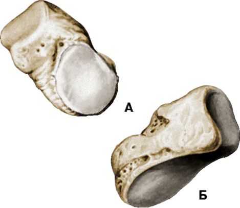 Рис. 96. Трехгранная кость, правая (А - ладонная поверхность, Б - тыльная поверхность)
 Рис. 97. Ладьевидная кость, правая (А - ладонная поверхность, Б - тыльная поверхность):
1 - Scaphoid, tubercle
 Рис. 98. Полулунная кость, правая (А - ладонная поверхность, Б - тыльная поверхность, В - дистальная поверхность)
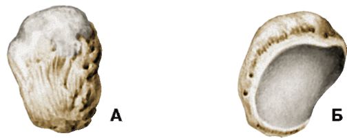 Рис. 99. Гороховидная кость, правая (А - ладонная поверхность, Б - тыльная поверхность)
 Рис. 100. Кость-трапеция, правая (А - ладонная поверхность, Б - тыльная поверхность):
1 - Trapezium, tubercle
 Рис. 101. Трапециевидная кость, правая (А - ладонная поверхность, Б - тыльная поверхность)
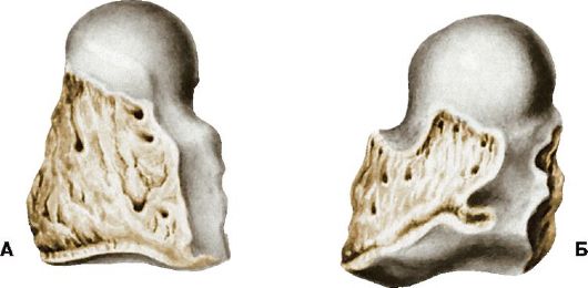 Рис. 102. Головчатая кость, правая (А - ладонная поверхность, Б - тыльная поверхность)
 Рис. 103. Крючковидная кость, правая (А - ладонная поверхность, Б - тыльная поверхность, В - вид снизу):
1 - Hook of hamate
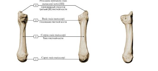 Головка пястной кости
АБ В
Рис. 104. Пястная кость [III], правая (А - ладонная поверхность, Б - тыльная поверхность, В - локтевая поверхность):
1 - Head of metacarpal; 2 - Shaft of metacarpal; Body of metacarpal; 3 - Base of metacarpal; 4 - Styloid process of third metacarpal [III]
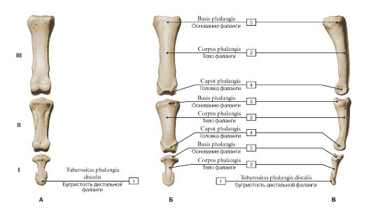 Рис.
105. Фаланги пальца правой кисти [III] (А - ладонная поверхность, Б -
тыльная поверхность, В - локтевая поверхность, I - проксимальная, II -
средняя, III - дистальная):
1 - Tuberosity of distal phalanx; 2 - Shaft of phalanx; Body of phalanx; 3 - Base of phalanx; 4 - Head of phalanx
 АБ
АБ
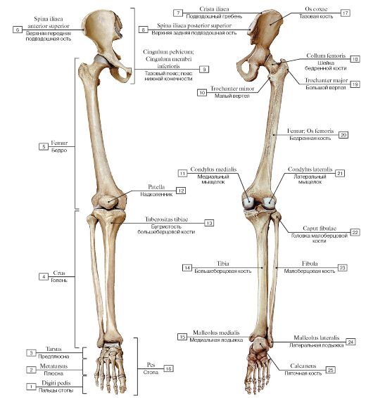
Рис. 106. Кости нижней конечности, правой (А - вид спереди, Б - вид сзади):
1 - Toes; 2 - Metatarsus; 3 - Ankle; 4 - Leg; 5 - Thigh; 6 - Anterior superior iliac spine; 7 - Iliac crest; 8 - Posterior superior iliac spine; 9 - Pelvic girdle; 10 - Lesser trochanter; 11 - Medial condyle; 12 - Patella; 13 - Tibial tuberosity; 14 - Tibia; 15 - Medial malleolus; 16 - Foot; 17 - Hip bone; Coxal bone; Pelvic bone; 18 - Neck of femur; 19 - Greater trochanter; 20 - Femur; Thigh bone; 21 - Lateral condyle; 22 - Head of fibula; 23 - Fibula; 24 - Lateral malleolus; 25 - Calcaneus
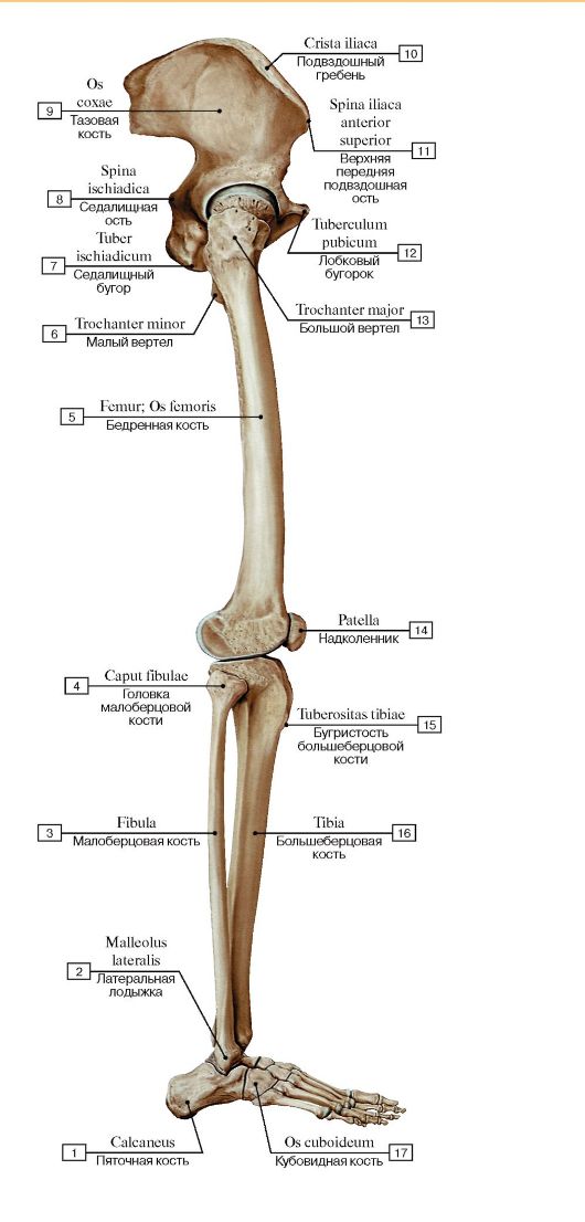 Рис. 107. Кости нижней конечности, правой, вид сбоку:
1 - Calcaneus; 2 - Lateral malleolus; 3 - Fibula; 4 - Head of fibula; 5 - Femur; Thigh bone; 6 - Lesser trochanter; 7 - Ischial tuberosity; 8 - Ischial spine; 9 - Hip bone; Coxal bone; Pelvic bone; 10 - Iliac crest; 11 - Anterior superior iliac spine; 12 - Pubic tubercle; 13 - Greater trochanter; 14 - Patella; 15 - Tibial tuberosity; 16 - Tibia; 17 - Cuboid
Рис. 108. Тазовая кость, правая (А - цветом выделены отдельные кости, Б - вид с латеральной стороны):
1 - Ischial tuberosity; 2 - Ischium, ramus; 3 - Ischial spine; 4 - Body of ischium; 5 - Ilium; 6 - Ala of ilium; Wing of ilium; 7 - Iliac crest; 8 - Acetabulum; 9 - Pubis, body; 10 - Superior pubic ramus; 11 - Inferior pubic ramus; 12 - Obturator foramen; 13 - Lesser sciatic notch; 14 - Greater sciatic notch; 15 - Posterior inferior iliac spine; 16 - Posterior superior iliac spine; 17 - Gluteal surface; 18 - Anterior gluteal line; 19 - Inferior gluteal line; 20 - Anterior superior iliac spine; 21 - Anterior inferior iliac spine; 22 - Acetabular margin; 23 - Lunate surface; 24 - Acetabular fossa; 25 - Acetabular notch; 26 - Pubic tubercle
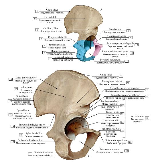
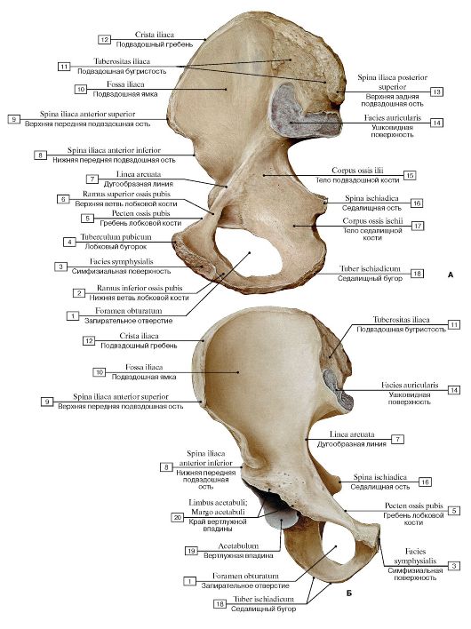 Рис. 109. Тазовая кость, правая (А - вид с медиальной стороны, Б - вид спереди):
1 - Obturator foramen; 2 - Inferior pubic ramus; 3 - Symphysial surface; 4 - Pubic tubercle; 5 - Pecten pubis; Pectineal line; 6 - Superior pubic ramus; 7 - Arcuate line; 8 - Anterior inferior iliac spine; 9 - Anterior superior iliac spine; 10 - Iliac fossa; 11 - Iliac tuberosity; 12 - Iliac crest; 13 - Posterior superior iliac spine; 14 - Ilium, auricular surface; 15 - Body of ilium; 16 - Ischial spine; 17 - Body of ischium; 18 - Ischial tuberosity; 19 - Acetabulum; 20 - Acetabular margin
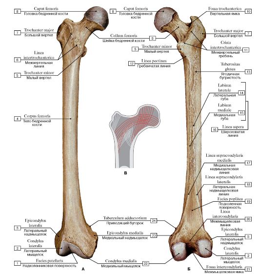 Рис.
110. Бедренная кость, правая (А - вид спереди, Б - вид сзади, В -
направления костных трабекул головки и шейки бедренной кости
относительно прилагаемой нагрузки):
1 - Patellar surface; 2 - Lateral condyle; 3 - Lateral epicondyle; 4 - Shaft of femur; Body of femur; 5 - Lesser trochanter; 6 - Intertrochanteric line; 7 - Greater trochanter; 8 - Head of femur; 9 - Neck of femur; 10 - Trochanteric fossa; 11 - Intertrochanteric crest; 12 - Pectineal line; Spiral line; 13 - Gluteal tuberosity; 14 - Lateral lip; 15 - Medial lip; 16 - Linea aspera; 17 - Medial supracondylar line; 18 - Lateral supracondylar line; 19 - Popliteal surface; 20 - Intercondylar line; 21 - Intercondylar fossa; 22 - Medial condyle; 23 - Medial epicondyle;
24 - Adductor tubercle
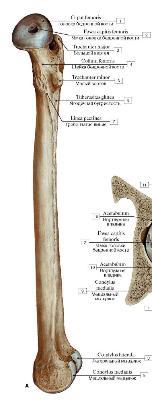 Рис. 112. Бедренная кость, правая (А - вид сбоку, с медиальной стороны, Б - верхний эпифиз):
1 - Head of femur; 2 - Fovea for ligament of head; 3 - Greater trochanter; 4 - Neck of femur; 5 - Lesser trochanter; 6 - Gluteal tuberosity; 7 - Pectineal line; Spiral line; 8 - Lateral condyle; 9 - Medial condyle; 10 - Acetabulum; 11 - Acetabular labrum; 12 - Patellar surface; 13 - Patella
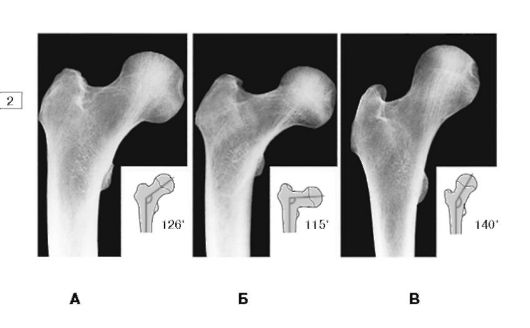 Рис.
111. Варианты соединения шейки с телом бедренной кости (А - нормальное
положение, Б - варусное положение, В - валыусное положение)
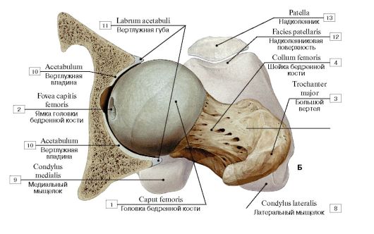
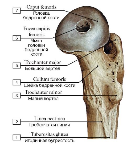 Рис. 113. Верхний эпифиз бедренной кости, правой, вид с медиальной стороны:
1 - Gluteal tuberosity; 2 - Pectineal line; Spiral line; 3 - Lesser trochanter; 4 - Neck of femur; 5 - Greater trochanter; 6 - Fovea for ligament of head; 7 - Head of femur
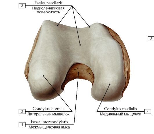 Рис. 114. Нижний эпифиз бедренной кости, правой,вид спереди:
1 - Intercondylar fossa; 2 - Lateral condyle; 3 - Patellar surface; 4 - Medial condyle
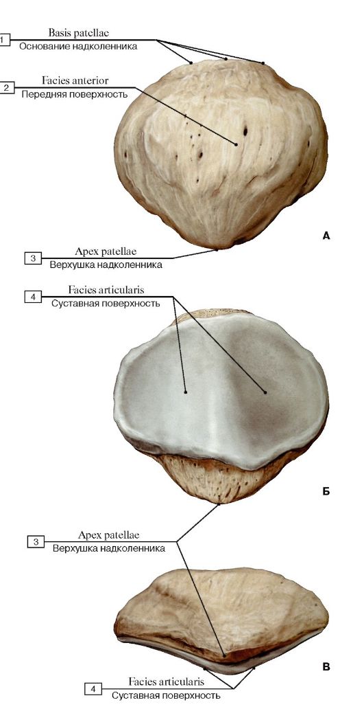 Рис. 115. Надколенник, правый (А - передняя поверхность, Б - суставная поверхность, В - вид сбоку)
1 - Base of patella; 2 - Anterior surface; 3 - Apex of patella; 4 - Articular surface
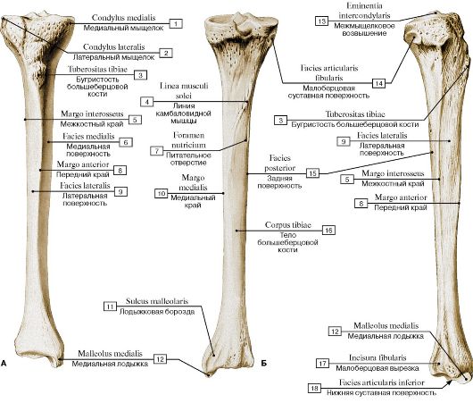
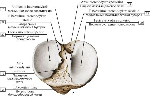 Рис.
116. Большеберцовая кость, правая (А - вид спереди, Б - вид сзади, В -
вид с латеральной стороны, Г - проксимальный эпифиз, вид сверху):
1 - Medial condyle; 2 - Lateral condyle; 3 - Tibial tuberosity; 4 - Soleal line; 5 - Interosseous border; 6 - Medial surface; 7 - Nutrient foramen; 8 - Anterior border; 9 - Lateral surface; 10 - Medial border; 11 - Malleolar groove; 12 - Medial malleolus; 13 - Intercondylar eminence; 14 - Fibular articular facet; 15 - Posterior surface; 16 - Shaft of tibia; Body of tibia; 17 - Fibular notch; 18 - Inferior articular surface; 19 - Anterior intercondylar area; 20 - Superior articular surface; 21 - Lateral intercondylar tubercle; 22 - Lateral intercondylar area; 23 - Medial intercondylar tubercle
В
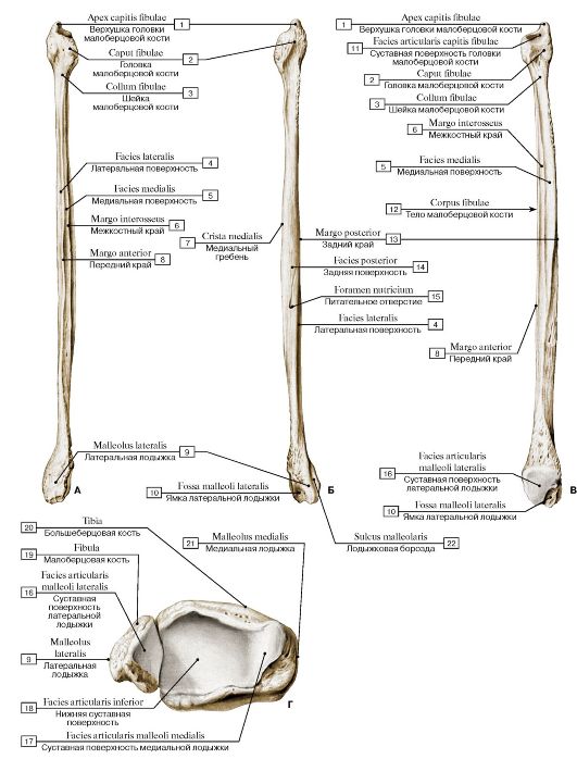 Рис.
117. Малоберцовая кость, правая (А - вид спереди; Б - вид сзади; В -
вид с медиальной стороны; Г - суставные поверхности нижних эпифизов
костей голени):
1 - Apex of head; 2 - Head of fibula; 3 - Neck of fibula; 4 - Lateral surface; 5 - Medial surface; 6 - Interosseous border; 7 - Medial crest; 8 - Anterior border; 9 - Lateral malleolus; 10 - Malleolar fossa; 11 - Articular facet; 12 - Shaft of fibula; Body of fibula; 13 - Posterior border; 14 - Posterior surface; 15 - Nutrient foramen; 16 - Articular facet; 17 - Articular facet; 18 - Inferior articular surface; 19 - Fibula; 20 - Tibia; 21 - Medial malleolus; 22 - Malleolar groove
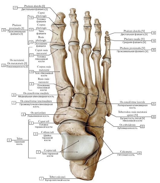 Рис. 118. Кости стопы, правой, вид сверху:
1 - Calcaneal tuberosity; 2 - Body of talus; 3 - Neck of talus; 4 - Head of talus; 5 - Talus; 6 - Navicular; 7 - Intermediate cuneiform; Middle cuneiform; 8 - Medial cuneiform; 9 - Base of metatarsal; 10 - Shaft of metatarsal; Body of metatarsal; 11 - Head of metatarsal; 12 - Metatarsal [I]; 13 - Base of phalanx; 14 - Shaft of phalanx; Body of phalanx; 15 - Head of phalanx; 16 - Proximal phalanx [I]; 17 - Distal phalanx [I]; 18 - Distal phalanx [V]; 19 - Middle phalanx [V]; 20 - Proximal phalanx [V]; 21 - Lateral cuneiform; 22 - Tuberosity of fifth metatarsal bone [V]; 23 - Cuboid; 24 - Calcaneus
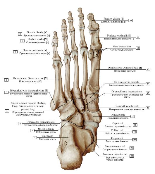 Рис. 119. Кости стопы, правой, вид снизу:
1 - Calcaneus; 2 - Cuboid; 3 - Tuberosity of cuboid; 4 - Groove for tendon of fibularis longus; Groove for tendon of peroneus longus; 5 - Tuberosity of first metatarsal bone [I]; 6 - Metatarsal [V]; 7 - Proximal phalanx [V]; 8 - Middle phalanx [V]; 9 - Distal phalanx [V]; 10 - Distal phalanx [I]; 11 - Proximal phalanx [I]; 12 - Sesamoid bones; 13 - Metatarsal [I]; 14 - Medial cuneiform; 15 - Intermediate cuneiform; Middle cuneiform; 16 - Lateral cuneiform; 17 - Navicular; 18 - Head of talus; 19 - Neck of talus; 20 - Body of talus; 21 - Sustentaculum tali; Talar shelf; 22 - Talus, posterior process
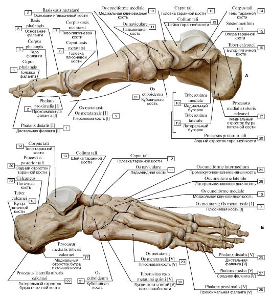 Рис. 120. Кости стопы, правой (А - вид с медиальной стороны, Б - вид с латеральной стороны):
I - Distal phalanx [I]; 2 - Proximal phalanx [I]; 3 - Head of phalanx; 4 - Shaft of phalanx; Body of phalanx; 5 - Base of phalanx; 6 - Head of metatarsal; 7 - Shaft of metatarsal; Body of metatarsal; 8 - Base of metatarsal; 9 - Metatarsal [I]; 10 - Medial cuneiform;
II - Navicular; 12 - Head of talus; 13 - Neck of talus; 14 - Body of talus; 15 - Sustentaculum tali; Talar shelf; 16 - Calcaneal tuberosity; 17 - Calcaneus, medial process; 18 - Medial tubercle; 19 - Lateral tubercle; 20 - Talus, posterior process; 21 - Cuboid; 22 - Calcaneus, lateral process; 23 - Calcaneus; 24 - Intermediate cuneiform; Middle cuneiform; 25 - Lateral cuneiform; 26 - Distal phalanx [V]; 27 - Middle
phalanx [V]; 28 - Proximal phalanx [V]; 29 - Metatarsal [V]; 30 - Tuberosity of fifth metatarsal bone [V]
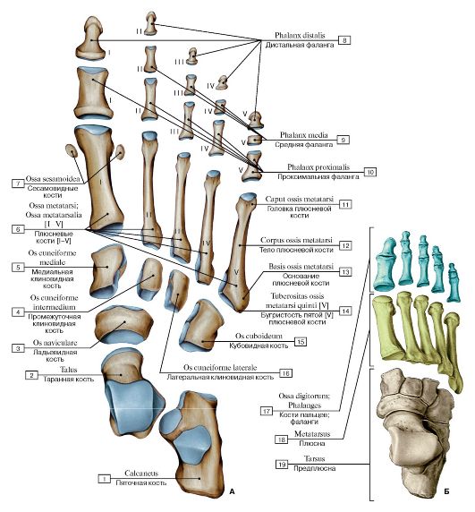 Рис. 121. Кости стопы, правой, вид сверху (А - кости, Б - отделы стопы):
1 - Calcaneus; 2 - Talus; 3 - Navicular; 4 - Intermediate cuneiform; Middle cuneiform; 5 - Medial cuneiform; 6 - Metatarsals [I-V]; 7 - Sesamoid bones; 8 - Distal phalanx; 9 - Middle phalanx; 10 - Proximal phalanx; 11 - Head of metatarsal; 12 - Shaft of metatarsal; Body of metatarsal; 13 - Base of metatarsal; 14 - Tuberosity of fifth metatarsal bone [V]; 15 - Cuboid; 16 - Lateral cuneiform; 17 - Phalanges;
18 - Metatarsus; 19 - Ankle
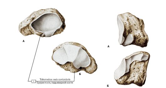 Рис. 122. Ладьевидная кость, правая (А - вид сзади, Б - вид спереди): Рис. 123. Медиальная клиновидная кость,
правая (А - медиальная поверхность,
1 - Navicular tuberosity Б - латеральная поверхность)
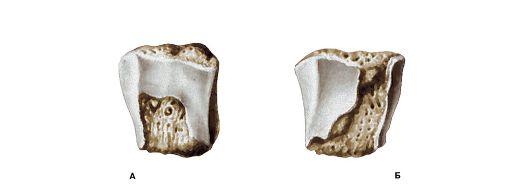 Рис. 124. Промежуточная клиновидная кость, правая (А - медиальная поверхность, Б - латеральная поверхность)
 Рис. 125. Латеральная клиновидная кость, правая (А - медиальная поверхность, Б - латеральная поверхность)
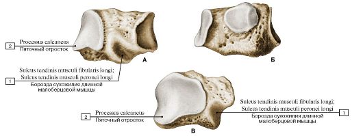 Рис. 126. Кубовидная кость, правая (А - латеральная поверхность, Б - медиальная поверхность, В - задняя
поверхность):
1 - Groove for tendon of fibularis longus; Groove for tendon of peroneus longus; 2 - Calcaneal process
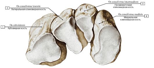 Рис. 127. Кости предплюсны, правые. Дистальный ряд:
1 - Cuboid; 2 - Lateral cuneiform; 3 - Intermediate cuneiform; Middle cuneiform; 4 - Medial cuneiform
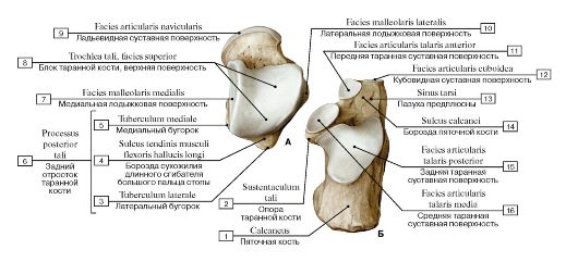 Рис. 128. Таранная (А) и пяточная (Б) кости, правые, вид сверху:
1 - Calcaneus; 2 - Sustentaculum tali; Talar shelf; 3 - Lateral tubercle; 4 - Groove for tendon of flexor hallucis longus; 5 - Medial tubercle; 6 = 3 + 4 + 5 - Posterior process; 7 - Medial malleolar facet; 8 - Trochlea of talus, superior facet; 9 - Navicular articular surface; 10 - Lateral malleolar facet; 11 - Anterior talar articular surface; 12 - Articular surface for cuboid; 13 - Tarsal sinus; 14 - Calcaneal sulcus; 15 - Posterior talar articular surface; 16 - Middle talar articular surface
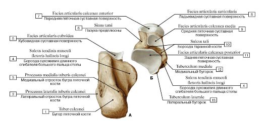 Рис. 129. Пяточная (А) и таранная (Б) кости, правые, вид снизу:
1 - Calcaneal tuberosity; 2 - Lateral process; 3 - Medial process; 4 - Groove for tendon of flexor hallucis longus; 5 - Articular surface for cuboid; 6 - Tarsal sinus; 7 - Anterior facet for calcaneus; 8 - Navicular articular surface; 9 - Middle facet for calcaneus; 10 - Sulcus tali; 11 - Posterior calcaneal articular facet; 12 - Medial tubercle; 13 - Lateral tubercle
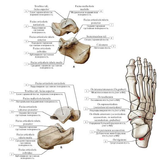 Рис. 131. Добавочные сесамовидные кости стопы, правой:
Рис. 130. Таранная и пяточная кости, правые (А - вид с медиальной стороны, Б - вид с латеральной стороны):
1 - Middle talar articular surface; 2 - Articular surface for cuboid; 3 - Anterior talar articular surface; 4 - Navicular articular surface; 5 - Trochlea of talus, superior facet; 6 - Medial malleolar facet; 7 - Posterior talar articular surface; 8 - Sustentaculum tali; Talar shelf; 9 - Calcaneus; 10 - Posterior calcaneal articular facet; 11 - Lateral malleolar facet
1 - Intermetatarsal bone; 2 - Vesalianum bone; 3 - Supranavicular bone; 4 - External tibial bone; 5 - Peroneal accessorial bone; 6 - Trigonal bone
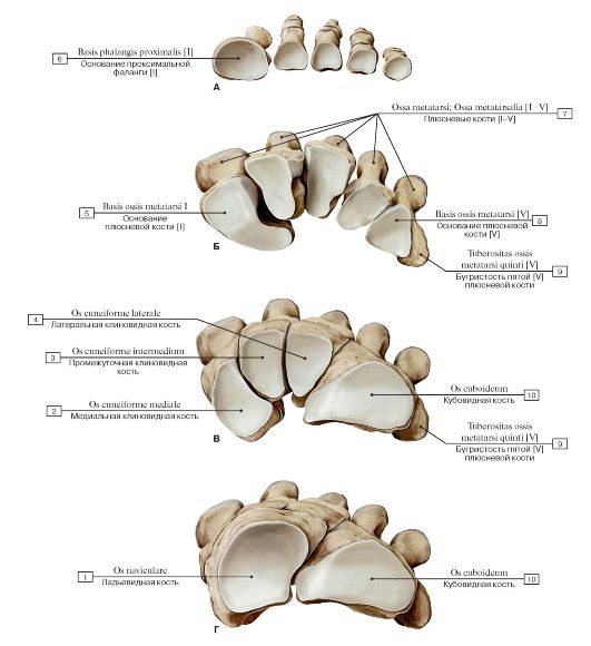 Рис.
132. Кости стопы, правой, вид сверху (А - основания проксимальных
фаланг, Б - основания костей плюсны, В - клиновидные и кубовидные кости,
Г - ладьевидная и кубовидная кости):
1 - Navicular; 2 - Medial cuneiform; 3 - Intermediate cuneiform; Middle cuneiform; 4 - Lateral cuneiform; 5 - Base of metatarsal [I]; 6 - Base of proximal phalanx [I]; 7 - Metatarsals [I-V]; 8 - Base of metatarsal [V]; 9 - Tuberosity of fifth metatarsal bone [V];
10 - Cuboid
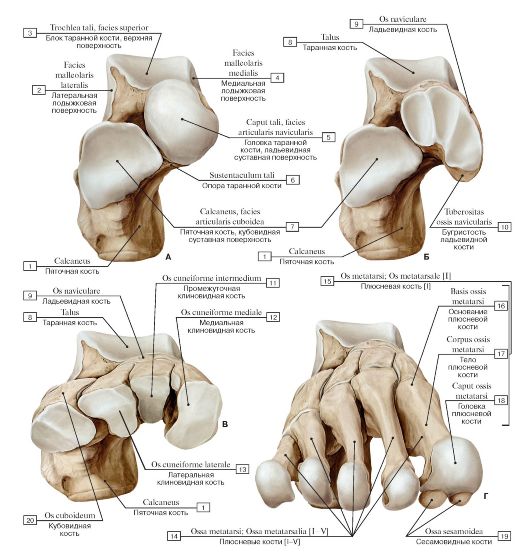 Рис.
133. Кости предплюсны и плюсны, правые (А - таранная и пяточная кости, Б
- таранная, пяточная и ладьевидная кости, В - таранная, пяточная,
ладьевидная и клиновидная кости, Г -кости плюсны, вид сверху и спереди):
1 - Calcaneus; 2 - Lateral malleolar facet; 3 - Trochlea of talus, superior facet; 4 - Medial malleolar facet; 5 - Head of talus, navicular articular surface; 6 - Sustentaculum tali; Talar shelf; 7 - Calcaneus, articular surface for cuboid; 8 - Talus; 9 - Navicular; 10 - Tuberosity; 11 - Intermediate cuneiform; Middle cuneiform; 12 - Medial cuneiform; 13 - Lateral cuneiform; 14 - Metatarsals [I-V]; 15 = 16 + 17 + 18 - Metatarsal I; 16 - Base of metatarsal; 17 - Shaft of metatarsal; Body of metatarsal; 18 - Head of metatarsal; 19 - Sesa-
moid bones; 20 - Cuboid
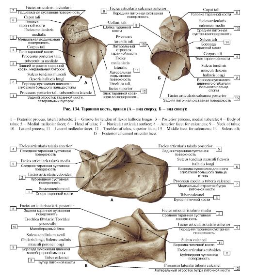 Рис. 135. Пяточная кость, правая (А - вид с медиальной стороны, Б - вид с латеральной стороны):
1 - Sustentaculum tali; Talar shelf; 2 - Articular surface for cuboid; 3 - Middle talar articular surface; 4 - Anterior talar articular surface; 5 - Posterior talar articular surface; 6 - Groove for tendon of flexor hallucis longus; 7 - Medial process; 8 - Calcaneal tuberosity; 9 - Groove for tendon of fibularis longus; Groove for tendon of peroneus longus; 10 - Fibular trochlea; Peroneal trochlea; Peroneal tubercle; 11 - Cal-
caneal sulcus; 12 - Lateral process
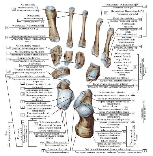 Рис. 136. Кости стопы, правой:
1 - Sustentaculum tali; Talar shelf; 2 = 3 + 7 + 8 + 9 + 10 + 11 + 12 + 13 - Talus; 3 = 4 + 5 + 6 - Posterior process; 4 - Lateral tubercle; 5 - Groove for tendon of flexor hallucis longus; 6 - Medial tubercle; 7 - Lateral process; 8 - Lateral malleolar facet; 9 - Trochlea of talus; 10 - Medial malleolar facet; 11 - Neck oftalus; 12 - Head oftalus; 13 - Navicular articular surface; 14 - Tuberosity; 15 - Navicular; 16 - Lateral cuneiform; 17 - Intermediate cuneiform; Middle cuneiform; 18 - Medial cuneiform; 19 - Tuberosity of first metatarsal bone [I]; 20 - Metatarsal [I]; 21 - Metatarsal [II]; 22 - Metatarsal [III]; 23 - Metatarsal [IV]; 24 = 25 + 26 + 27 + 28 - Metatarsal [V]; 25 - Head of metatarsal; 26 - Shaft of metatarsal; Body of metatarsal; 27 - Base of metatarsal; 28 - Tuberosity of fifth metatarsal bone [V]; 29 = 30 + 31 + 32 - Cuboid; 30 - Groove for tendon of fibularis longus; Groove for tendon of peroneus longus; 31 - Tuberosity; 32 - Anterior facet for calcaneus; 33 = 34 + 35 + 36 + 37 + 38 + 39 - Calcaneus; 34 - Articular surface for cuboid; 35 - Anterior talar articular surface; 36 - Calcaneal sulcus; 37 - Middle talar articular surface; 38 - Posterior talar articular surface; 39 = 40 + 41 - Calcaneal tuberosity; 40 - Medial process; 41 - Lateral process
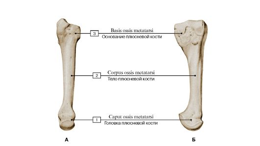 Рис. 137. Плюсневая кость [III], правая (А - подошвенная поверхность, Б - локтевая поверхность):
1 - Head of metatarsal; 2 - Shaft of metatarsal; Body of metatarsal; 3 - Base of metatarsal
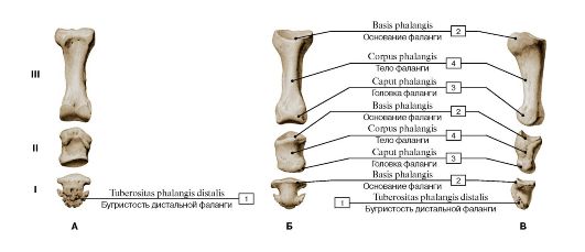 Рис.
138. Фаланги пальца стопы [III], правой (А - тыльная поверхность, Б -
подошвенная поверхность, В - латеральная поверхность, I - проксимальная,
II - средняя, III - дистальная):
1 - Tuberosity of distal phalanx; 2 - Base of phalanx; 3 - Head of phalanx; 4 - Shaft of phalanx; Body of phalanx